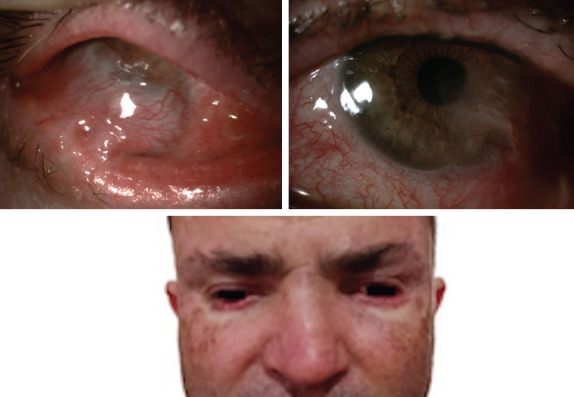
Figure 1.

This picture depicts anterior segment photography of both eyes. Symblepharon formation was observed extending from the tarsal conjunctiva to the bulbar conjunctiva and spreading over the cornea, preventing the selection of the anterior chamber in the right eye (upper left). The left eye image shows more apparent anterior chamber detail than the right eye. Symblepharon formation extended from the inferior eyelid and inferior bulbar conjunctiva to the temporal cornea (upper right). Pigmented lesions were observed in the maxillofacial–temporal areas of the patient’s face. Upon anamnesis, it was learned that basal cell carcinoma-nodular type was diagnosed due to the biopsy taken from the pigmented lesions on the skin (bottom).
