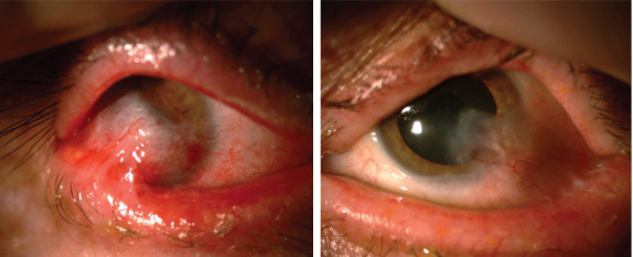
Figure 3.

Appearance after ocular surface rehabilitation surgery. The aggressive symblepharon formation in the right eye has regressed, and the anterior chamber details have become more visible (left image). In the left eye, ocular surface findings remained the same (right image).
