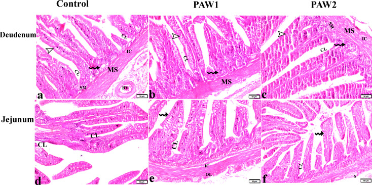
Fig. 2.
The effects of PAW on duodenum and jejunum histopathological alterations in quail. (a-c): duodenum tissue sections from experimental groups. villi (V), mucosa (M), submucosa (SM), muscularis (MS), serosa (S) and blood vessel (Bv), inner circular (IC) and the outer longitudinal (OL) muscularis externa, simple columnar epithelial cells with goblet cells (arrowheads), crypts of Lieberkühn (CL), glands of Lieberkühn lined with Paneth cells (zigzag arrows). (d-f): jejunum tissue sections from experimental groups. villi (V), simple columnar epithelial cells with goblet cells (Zigzag arros), crypts of Lieberkuhn (CL), glands of Lieberkuhn (GL), muscularis mucosa (MM), muscularis externa; inner circular muscle (IC), outer longitudinal muscle (OL), serosa (S). There is no structural difference between the groups

