Fig. 5.
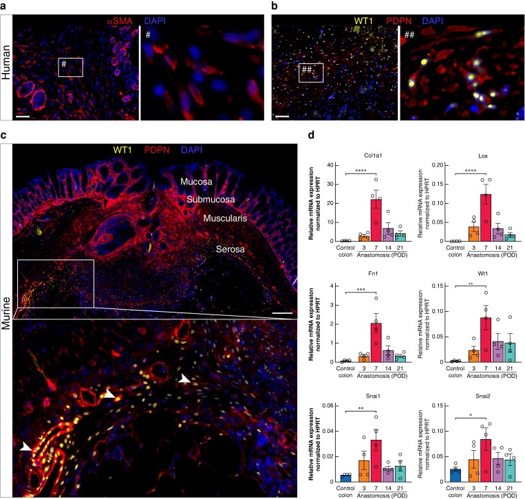
Cells within the anastomotic serosal scar express mesothelial cell markers
a Immunofluorescence (IF) staining for alpha smooth muscle actin (αSMA) shows αSMA+ cells and microvessels within the serosal scar of human anastomosis at postoperative day (POD) 7. Scale bar = 50 µm, 200× magnification. b IF staining for Wilms tumour protein (WT1) and podoplanin (PDPN) showing the presence of WT1+PDPN+ mesothelial cells (MCs) within the anastomotic scar of human anastomosis at POD 7. Scale bar = 50 µm 200× magnification. c IF staining for WT1 and PDPN showing the presence of WT1+PDPN+ MCs within the anastomotic scar of murine anastomosis at POD 7. White arrowheads: transition of WT1+PDPN+ cells from epithelial to mesenchymal phenotype within the anastomotic scar. Scale bar = 200 µm, 100× magnification, n = 3. d Relative gene expression of extracellular matrix-associated proteins (Col1a1, Fn1, Lox), mesothelial cell markers (Wt1) and key epithelial-to-mesenchymal transition (EMT) transcription factors (Snai1, Snai2) within murine anastomosis at POD 3, 7, 14 and 21 compared to murine control colon. n = 4 per time point, one-way ANOVA with Tukey’s multiple comparison test to control colon, *P < 0.05, **P < 0.01, ***P < 0.001, ****P < 0.0001. Data are mean(s.e.m.) with dots for individual values.

