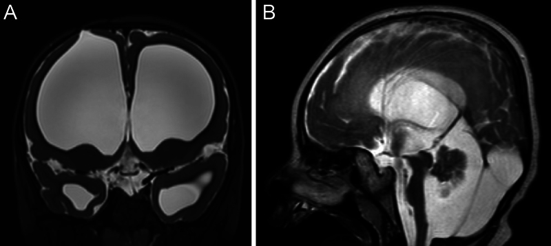
FIG. 1.
Preoperative coronal (A) and sagittal (B) T2-weighted magnetic resonance imaging of the brain. Coronal imaging demonstrates the bony and cortical entry point of the old ETV. Note the pronounced ventriculomegaly and thin cortex. The corpus callosum is so thin that it is not readily appreciable in the sagittal plane.

