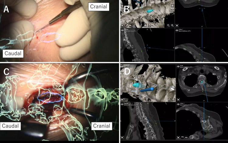
FIG. 3.
Intraoperative microscopic views with AR objects and navigation maps during the approach. The green object represents the bone, and the blue object represents the syrinx. A: Skin incision. B: Navigation map during skin incision. C: Exposure of the lamina. D: Navigation map during exposure of the lamina.

