Figure 3.

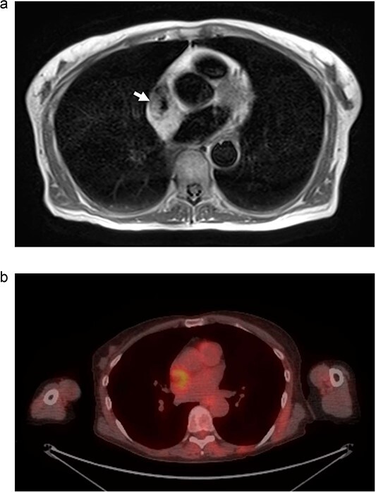
(a) MRI chest. Mass represented by white arrow. (b) PET/CT. Uptake is demonstrated in the mass associated with the SVC and right atrium.

(a) MRI chest. Mass represented by white arrow. (b) PET/CT. Uptake is demonstrated in the mass associated with the SVC and right atrium.