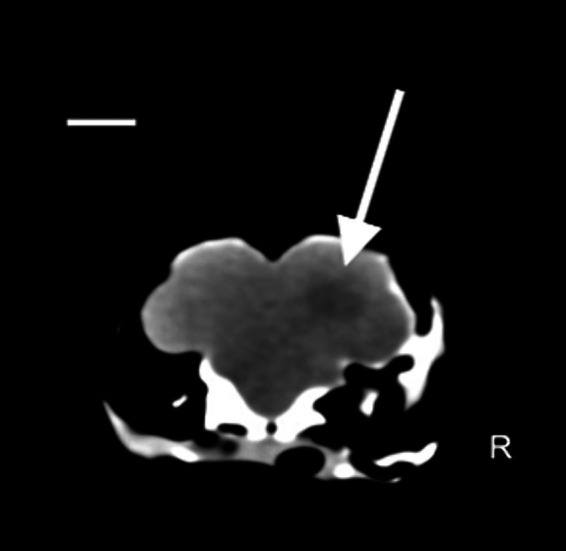
Figure 2.

Coronal plane image with soft tissue algorithm in the brain window. Scale bar = 8 mm. A hypoattenuating ovalar intra-axial lesion with ill-defined margin is observed at level of parieto-temporal area at the level of the right cerebral hemisphere (white arrow) (Pitch 0.562, slice thickness: 1.25 mm).
