Figure 6.
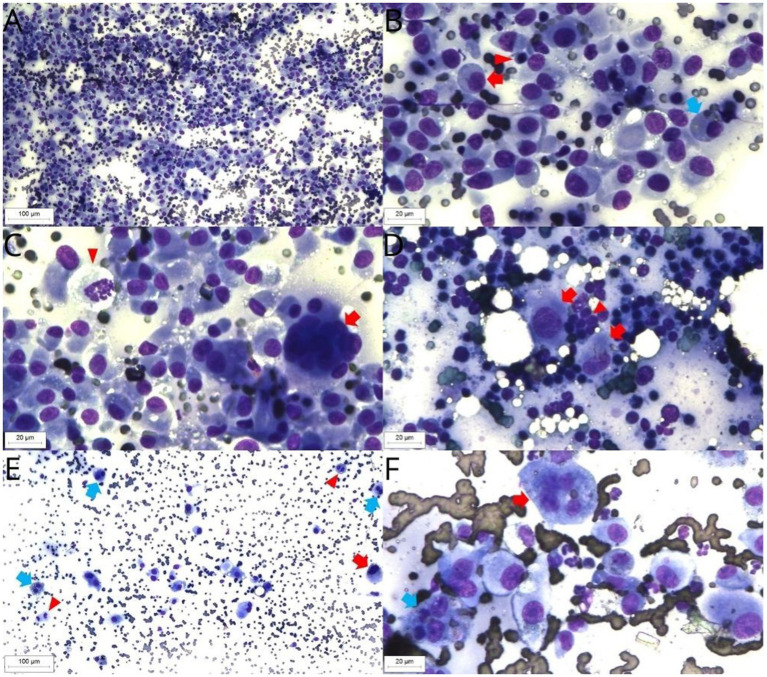
FPH cytology, Diff Quick staining. Retropharyngeal lymph node sample. Background with moderate red blood cells numbers. High large histiocytic cells, moderate small lymphocytes and low neutrophils numbers (10× objective) (A). Retropharyngeal lymph node sample. Histiocytic cells have a moderate nucleus-to-cytoplasm ratio, rounded or oval nucleus, moderate anisokaryosis, single nucleolus and low to moderate basophilic cytoplasm (red arrow). Presence of cytophagocytosis (blue arrow). The small lymphocytes present typical morphology (arrowhead) (40× objective) (B). Retropharyngeal lymph node. Multinucleated histiocytic cell (arrow) and mitotic figure (arrowhead) (40× objective) (C). Popliteal lymph node sample. Presence of high small lymphocytes (arrowhead) and a low histiocytic cells numbers, with the last ones exhibiting the same pattern observed in the retropharyngeal lymph node sample (red arrow) (40× objective) (D). Superficial cervical lymph node. Background with high red blood cells numbers. Presence of large histiocytic cells, showing high anisokaryosis (red arrow), binucleated cells (blue arrow) and mitotic figures (arrowhead) (10× objective) (E). Superficial cervical lymph node sample. Histiocytic cells with a prominent single nucleolus, binucleation with cytophagocytosis (blue arrow) and multinucleation (red arrow) (40× objective) (F).

