Abstract
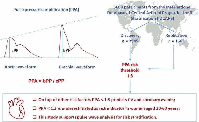
Pulse pressure amplification (PPA) is the brachial-to-aortic pulse pressure ratio and decreases with age and cardiovascular risk factors. This individual-participant meta-analysis of population studies aimed to define an outcome-driven threshold for PPA. Incidence rates and standardized multivariable-adjusted hazard ratios (HRs) of cardiovascular and coronary endpoints associated with PPA, as assessed by the SphygmoCor software, were evaluated in the International Database of Central Arterial Properties for Risk Stratification (n = 5608). Model refinement was assessed by the integrated discrimination (IDI) and net reclassification (NRI) improvement. Age ranged from 30 to 96 years (median 53.6). Over 4.1 years (median), 255 and 109 participants experienced a cardiovascular or coronary endpoint. In a randomly defined discovery subset of 3945 individuals, the rounded risk-carrying PPA thresholds converged at 1.3. The HRs for cardiovascular and coronary endpoints contrasting PPA < 1.3 vs ≥1.3 were 1.54 (95% confidence interval [CI]: 1.00–2.36) and 2.45 (CI: 1.20–5.01), respectively. Models were well calibrated, findings were replicated in the remaining 1663 individuals analyzed as test dataset, and NRI was significant for both endpoints. The HRs associating cardiovascular and coronary endpoints per PPA threshold in individuals <60 vs ≥60 years were 3.86 vs 1.19 and 6.21 vs 1.77, respectively. The proportion of high-risk women (PPA < 1.3) was higher at younger age (<60 vs ≥60 years: 67.7% vs 61.5%; P < 0.001). In conclusion, over and beyond common risk factors, a brachial-to-central PP ratio of <1.3 is a forerunner of cardiovascular coronary complications and is an underestimated risk factor in women aged 30–60 years. Our study supports pulse wave analysis for risk stratification.
Keywords: Pulse pressure amplification, Waveform analysis, Cardiovascular risk, Population science
Introduction
Pulse pressure (PP), the difference between systolic and diastolic blood pressure (BP), oscillates around mean arterial pressure and increases from the central to the peripheral arteries, causing PP amplification (PPA) [1–5]. PPA is most commonly represented by the ratio of brachial-to-aortic PP [1–5]. Over the human lifespan, PPA decreases as a consequence of arterial stiffening associated with aging and further promoted by hypertension, dyslipidemia, diabetes, and chronic kidney disease [4, 6]. Stiffening of the central elastic arteries leads to the earlier return of backward waves from peripheral reflection sites [1, 5], thereby increasing central systolic pressure, so that the ratio of brachial-to-aortic PP decreases. Among healthy, normotensive individuals enrolled in the Anglo-Cardiff Collaborative Trial (ACCT), PPA decreased from 1.72 in teenagers to 1.25 in octogenarians [7]. In populations [2] and patients [8], arterial stiffening, as captured by lower PPA, is associated with higher cardiovascular risk [2, 3], coronary artery diseases [9], deterioration of renal function [8], and mortality [3]. However, few prospective studies focused on the incidence of major cardiovascular complications in relation to PPA. To the best of our knowledge, there is therefore no published outcome-driven PPA threshold, which might alert clinicians for the risk of cardiovascular complications related to falling PPA. To address this issue, the current individual-participant level meta-analysis defined, calibrated and validated an outcome-driven threshold for PPA, using the International Database of Central Arterial Properties for Risk Stratification (IDCARS) [10].
Methods
Study participants
The study protocol paper describes the construction of the IDCARS database [10]. The longitudinal studies extracted from the IDCARS resource qualified for the current analysis, if information on brachial and central BP and cardiovascular risk factors was available at baseline, if the arterial pulse waveform had been measured, if follow-up included both fatal and nonfatal endpoints, if study reports were published in peer-reviewed journals, and if the study participants were representative for a population. All studies complied with the Helsinki Declaration on research in humans [11] and were approved by the competent Institutional Review Boards. Participants provided written informed consent. All data were stripped from personal identifiers, and if required by national legislations, additional ethical clearance was obtained. Supplementary Table S1 lists cohort-specific information on the catchment area, sampling strategies, timeframes of recruitment and follow-up, and initial participation rates. Enrolment took place from 1985 until 2015. For the present analysis, baseline refers to the first measurement of central and peripheral BP along with cardiovascular risk factors (October 2000 until February 2016). Across studies, the last follow-up took place from October 2012 to December 2018 (Supplementary Table S1). Of 6650 IDCARS participants qualifying for analysis, we excluded 1042 because they were younger than 30 years without endpoints (n = 954), peripheral PP was >130 mmHg (n = 10), central systolic BP was <70 mmHg (n = 1) or >230 mmHg (n = 1), central diastolic BP was >150 mmHg (n = 1) or <55 mmHg (n = 15), or because the pulse wave analysis was missing (n = 60). This left 5608 participants for statistical analysis.
Pulse wave analysis
Brachial BP, measured immediately prior to the hemodynamic assessment after participants had rested for 5 up to 15 min in the supine position, was the average of two consecutive readings. Experienced observers recorded the radial arterial waveform at the dominant arm during an 8-s period by applanation tonometry. They used a high-fidelity SPC-301 micromanometer (Millar Instruments Inc., Houston, TX), interfaced with a SphygmoCor CvMS device and a laptop computer running SphygmoCor software (AtCor Medical Inc., Itasca, IL). Recordings were discarded, if systolic or diastolic variability of consecutive waveforms exceeded 5% or if the amplitude of the pulse wave signal was below 80 mV, or if the operator index was <70%. From the radial signal, the SphygmoCor software reconstructs the aortic pulse wave by means of a validated generalized transfer function [12]. Estimates of central blood pressure were calibrated on brachial systolic and diastolic BP. PPA was the brachial-to-aortic PP ratio.
Ascertainment of endpoints
We ascertained the incidence of fatal and nonfatal endpoints from the appropriate sources in each country. The primary endpoint was a composite cardiovascular outcome consisting of cardiovascular mortality, sudden death and nonfatal endpoints, including myocardial infarction, heart failure, stroke, and coronary arterial revascularization. The coronary endpoint comprised sudden death and fatal and nonfatal myocardial infarction and coronary revascularization. The endpoints are listed in Supplementary Table S2. All endpoints were validated against hospital files or medical records held by primary care physicians or specialists. Only the first event within each category was considered in the analysis of outcome.
Statistical analysis
Statistical methods are described in detail in the online-only Data Supplement (pp. 2–5). In exploratory analyses, incidence rates of endpoints were tabulated by tertiles of the PPA distribution, while applying the direct method for standardizing rates for cohort, sex and age (<40, 40–59, ≥60 years). The cumulative incidence of the cardiovascular and coronary endpoints was plotted, while accounting for cohort, sex, age and heart rate.
Multivariable-adjusted Cox models accounted for cohort (random effect), sex, age, heart rate, body mass index, smoking and drinking, the total-to-HDL serum cholesterol ratio, the glomerular filtration rate estimated from serum creatinine by the chronic kidney disease epidemiology collaboration equation [13], antihypertensive drug intake, history of cardiovascular disease and diabetes. The proportional hazards assumption was checked by the Kolmogorov-type supremum test.
After stratification for sex, median age (53.6 years) and cohorts (n = 9; Supplementary Table S1), a random function was applied to subdivide the total IDCARS study population (n = 5608) into a discovery (n = 3945) and replication (n = 1663) sample. The size of the discovery dataset required 171 cardiovascular endpoints to demonstrate a difference between high-risk (above PPA risk threshold) and low-risk (below PPA risk thresholds) individuals with the α-level and power set at 0.05 and 0.80, respectively [14]. The default significance throughout the current study was a two-tailed α-level of ≤0.05 with the SE to compute two-sided confidence intervals [CI] set at 1.96. However, given the prior probability in the discovery dataset, in the replication analysis, the α-level was a one-tailed level of ≤0.05 and the SE to compute CIs was 1.65.
To determine an operational threshold for PPA in the discovery dataset, a two-pronged strategy [15, 16] was applied using Cox regression. First, multivariable-adjusted HRs were computed for 0.1 increments in PPA from the 10th to the 90th percentile of the PPA distribution. These HRs expressed the risk in participants, whose PPA exceeded the cut-off point vs the average risk in the whole population. The HRs with CIs were plotted as function of increasing PPA thresholds to assess at which PPA level the upper 95% confidence limit of the HRs crossed unity, signifying decreased risk [15]. Next, PPA thresholds were obtained by determining the PPA levels yielding a 5-year risk equivalent to the risk associated with a brachial systolic BP of 120-, 130-, 140- and 160 mmHg [16]. Model calibration was evaluated by comparing the predicted risk against overoptimism-corrected Kaplan–Meier estimates in PPA quintiles. The performance of PPA in risk stratification was assessed from 2-by-2 tables providing specificity, sensitivity and related statistics, the area (AUC) under the receiver operating curve (ROC), the area (AUC) under the receiver operating curve, and by the integrated discrimination improvement (IDI) and the net reclassification improvement (NRI) [17].
Finally, subgroup analyses were conducted in participants stratified by sex, age (<60 vs ≥60 years), median systolic BP (<130 vs ≥130 mmHg) and antihypertensive treatment status. To compare relative risk across these strata, deviation from mean coding [18] was applied. In a further sensitivity analysis, models for the coronary endpoint were additionally adjusted for diastolic BP, the driving force of the coronary circulation.
Results
Baseline characteristics of participants
Table 1 lists the main characteristics of the 5608 analyzed participants. Mean age at baseline was 54.2 years. Among all participants, 2988 (53.3%) had hypertension, 1946 (34.7%) were on antihypertensive treatment. Of 1946 patients reporting information on antihypertensive drugs, 672 (34.5%) and 1274 (65.5%) were taking a single agent or combination therapy, respectively. Drug classes taken were diuretics in 648 (33.3%) patients, β-blockers in 747 (38.4%), inhibitors of the renin–angiotensin system in 1184 (60.8%), and vasodilators in 833 (42.8%). Among the 5608 study participants (Table 1), 3035 (54.1%) were women. The PPA distributions is shown in Supplementary Fig. S1. The association between PPA and age is shown in Supplementary Fig. S2. Compared to women, more men reported smoking cigarettes (33.7% vs 10.2%) or consuming alcoholic beverages (75.4% vs 28.9%).
Table 1.
Baseline characteristics of participants
| Characteristic | Discovery | Replication | All |
|---|---|---|---|
| Number in group | 3945 | 1663 | 5608 |
| Number with characteristic (%) | |||
| Ethnicity | |||
| White Europeans | 1684 (42.7) | 701 (42.2) | 2385 (42.5) |
| Chinese | 1279 (32.4) | 544 (32.7) | 1823 (32.5) |
| South Americans | 982 (24.9) | 418 (25.1) | 1400 (25.0) |
| Women | 2134 (54.1) | 901 (54.2) | 3035 (54.1) |
| Hypertension | 2076 (52.6) | 912 (54.8) | 2988 (53.3) |
| Treated hypertension | 1346 (34.1) | 600 (36.1) | 1946 (34.7) |
| Diabetes mellitus | 246 (6.2) | 93 (5.6) | 339 (6.0) |
| History of cardiovascular disease | 555 (14.1) | 238 (14.3) | 793 (14.1) |
| Current smoking | 853 (21.6) | 326 (19.6) | 1179 (21.0) |
| Drinking alcohol | 1990 (50.4) | 826 (49.7) | 2816 (50.2) |
| Mean of characteristic (SD) | |||
| Age, y | 54.2 (14.5) | 54.2 (14.1) | 54.2 (14.4) |
| Body mass index, kg/m2 | 25.8 (4.9) | 25.9 (4.8) | 25.8 (4.8) |
| Brachial blood pressure, mmHg | |||
| Systolic | 134.1 (20.9) | 134.1 (21.2) | 134.1 (21.0) |
| Diastolic | 80.0 (10.6) | 80.5 (10.8) | 80.1 (10.7) |
| Pulse pressure | 54.1 (16.3) | 53.6 (16.3) | 53.9 (16.3) |
| Central blood pressure, mmHg | |||
| Systolic | 123.7 (21.0) | 123.9 (21.5) | 123.7 (21.2) |
| Diastolic | 81.0 (10.8) | 81.5 (11.0) | 81.2 (10.9) |
| Pulse pressure | 42.6 (16.0) | 42.4 (16.2) | 42.6 (16.1) |
| Heart rate, beats per min | 65.5 (11.4) | 65.6 (11.5) | 65.5 (11.4) |
| Total serum cholesterol, mg/dL | 194.8 (38.6) | 196.8 (39.8) | 195.4 (38.9) |
| HDL serum cholesterol, mg/dL | 57.4 (15.2) | 57.6 (15.2) | 57.5 (15.2) |
| Total-to-HDL cholesterol ratio | 3.6 (1.1) | 3.6 (1.1) | 3.6 (1.1) |
| Glomerular filtration rate (mL/min/1.73 m2) | 82.4 (19.9) | 82.6 (19.1) | 82.5 (19.6) |
| Blood glucose, mg/dL | 90.6 (18.8) | 90.9 (20.2) | 90.7 (19.2) |
| Pulse pressure amplification | 1.31 (0.19) | 1.31 (0.19) | 1.26 (1.16–1.42) |
| Median follow-up (IQR), years | 4.1 (3.6–6.9) | 4.2 (3.6–7.5) | 4.1 (3.6–6.9) |
After stratification for sex, median age (53.6 years) and cohorts, a random function was applied to subdivide the total IDCARS study population into a discovery and replication sample, which were well matched (P ≥ 0.09 for all characteristics). Current smoking was inhaling tobacco smoke on a daily basis. Drinking alcohol was the occasional or daily consumption of alcoholic beverages. Diabetes mellitus was use of anti-diabetic drugs, fasting blood glucose ≥126 mg/dL, random blood glucose ≥200 mg/dL, a self-reported diagnosis, or diabetes documented in practice or hospital records. Hypertension was a brachial blood pressure of ≥140 mmHg systolic or ≥90 mmHg diastolic, or use of antihypertensive drugs. The glomerular filtration rate was derived from serum creatinine using the chronic kidney disease epidemiology collaboration formula
HDL high-density lipoprotein
Incidence of endpoints
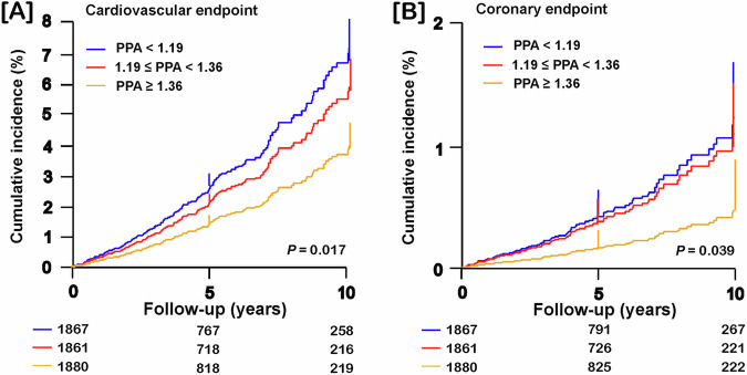
Over a median follow-up of 4.1 years (interquartile range (IQR): 3.6–6.9 years; 5th–95th percentile interval: 2.2–12.1 years), of 5608 participants, 255 (4.55%) experienced a cardiovascular endpoint and 109 (1.94%) a coronary endpoint, resulting in rates of 9.59 events per 1000 person-years (confidence interval (CI): 8.82–10.4) and 4.05 events per 1000 person-years (CI: 3.53–4.57), respectively. Across tertiles of the PPA distribution, the crude rates of cardiovascular and coronary endpoints decreased with higher PPA category (Supplementary Table S3). Figure 1 shows the cumulative incidence of the cardiovascular and coronary endpoints by PPA tertiles with adjustment for cohort (random effect), sex and age. Given that in the whole study population, PPA was a risk factor, in the next step of the analysis, we first determined an outcome-driven threshold in the discovery sample.
Fig. 1.
Cumulative incidence of the cardiovascular and coronary endpoints by tertiles of pulse pressure amplification. Cumulative incidence of the cardiovascular (A) and coronary (B) endpoints was derived by Cox regression with adjustment for cohort, sex and age. P values denote the overall significance of the difference between the PPA categories. Vertical lines denote the SE. Tabulated data are the number of participants at risk at 5-year intervals
Analysis of the discovery sample
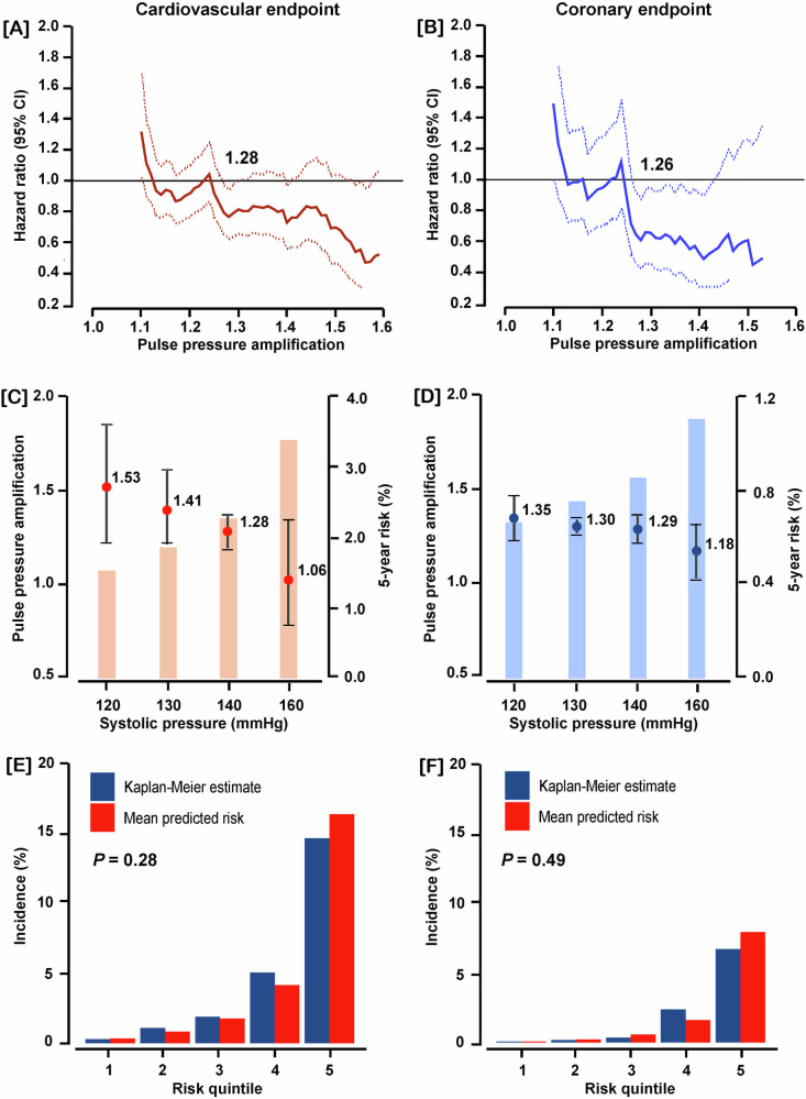
Multivariable-adjusted HRs were plotted against PPA thresholds stepwise increasing by 0.1 over the 10th–90th percentile range of the PPA distribution (Fig. 2A, B). These multivariable-adjusted HRs expressed the 5-year risks of cardiovascular and coronary endpoints associated with successively increasing PPA thresholds compared to the average risk in the discovery cohort. The upper 95% confidence limit of the HRs crossed unity at a PPA level of 1.28 and 1.26 for the cardiovascular and coronary endpoints, respectively. Furthermore, the PPA thresholds yielding a risk equivalent with a systolic BP of 140 mmHg were 1.28 (CI: 1.19–1.36) and 1.29 (CI: 1.22–1.36) for the cardiovascular and coronary endpoints (Fig. 2C, D). With rounding applied, the PPA thresholds distinguishing between low and high risk of the two endpoints endpoint were <1.3 vs ≥1.3). In the discovery dataset, the adjusted HRs contrasting PPA levels of <1.3 vs ≥1.3 were 1.54 (CI: 1.00–2.36) for the cardiovascular endpoint and 2.45 (CI: 1.20–5.01) for the coronary endpoint (Table 2). These models were well calibrated (Fig. 2E, F). A 1-SD PPA increment in the discovery cohort yielded multivariable-adjusted HRs of 0.74 (CI: 0.26–0.97) and 0.57 (CI: 0.36–0.91) for the cardiovascular and coronary endpoint (Table 2).
Fig. 2.
Threshold and calibration of pulse pressure amplification (PPA) in the discovery sample including 3945 participants. Hazard ratios (HRs) express the risk at each PPA level relative to the average risk in the whole discovery sample for cardiovascular (A) and coronary endpoints (B). The upper confidence limit crosses unity, denoted by the vertical line, at 1.28 and 1.26 for cardiovascular and coronary endpoints, signifying decreased risk. The PPA levels yielding equivalent 5-year risks compared with a systolic blood pressure of 140 mmHg were 1.28 and 1.29 for the cardiovascular (C) and coronary (D) endpoints, respectively. Model calibration for the cardiovascular (E) and coronary (F) endpoints demonstrated that across PPA quintiles the predicted risk was similar compared with the overoptimism-corrected Kaplan–Meier estimates. All analyses were multivariable adjusted for cohort (random effect), sex, age, body mass index, heart rate, smoking and drinking, total-to-HDL serum cholesterol ratio, estimated glomerular filtration rate, antihypertensive drug intake, history of cardiovascular disease, and diabetes
Table 2.
Endpoints in relation to PPA per threshold or analyzed as continuously distributed variable
| Cohort Endpoint Risk indicator | Ne/Nr | HR (95% CI) | P value |
|---|---|---|---|
| Discovery (n = 3945) | |||
| Cardiovascular | |||
| PPA < 1.3 vs ≥1.3 | 150/2230 vs 39/1715 | 1.54 (1.00–2.36) | 0.048 |
| PPA + 1 SD | 189/3945 | 0.74 (0.56–0.97) | 0.031 |
| Coronary | |||
| PPA < 1.3 vs ≥1.3 | 66/2230 vs 12/1715 | 2.45 (1.20–5.01) | 0.014 |
| PPA + 1 SD | 78/3945 | 0.57 (0.36–0.91) | 0.018 |
| Replication (n = 1663) | |||
| Cardiovascular | |||
| PPA < 1.3 vs ≥1.3 | 53/949 vs 13/714 | 2.02 (1.07–3.78) | 0.033 |
| PPA + 1 SD | 66/1663 | 0.61 (0.39–0.96) | 0.036 |
| Coronary | |||
| PPA < 1.3 vs ≥1.3 | 25/949 vs 6/714 | 2.90 (1.18–7.12) | 0.025 |
| PPA + 1 SD | 31/1663 | 0.61 (0.34–1.11) | 0.085 |
Models were adjusted for cohort (random effect), sex, age, body mass index, heart rate, smoking and drinking, total-to-HDL serum cholesterol ratio, estimated glomerular filtration rate, antihypertensive drug intake, history of cardiovascular disease, and diabetes. Given the prior probability generated in the discovery sample, confidence interval and P values in the replication sample are one-sided
Ne/Nr number of events/of participants at risk
Replication
In the replication population (Table 2), the HRs were similar, but given the prior probability generated in the discovery sample HRs are given with one-sided P values and confidence intervals. However, given the smaller sample size, significance was not reached for the coronary endpoint in the continuous analysis (P = 0.085).
Predictive performance
Supplementary Table S4 lists the performance of the 1.3 PPA threshold in the 5-year risk stratification and the AUC for the continuously distributed PPA. For the cardiovascular and coronary endpoints, the classification estimates derived from 2-by-2 tables were broadly similar in the discovery, replication and the whole dataset with specificity ~0.40 and sensitivity ~0.80. For the continuously distributed PPA, the AUC was around 0.60.
In the discovery and replication sample and in all participants, NRI for the 1.3 PPA threshold was significant, ranging from 16.1 to 33.7% and from 30.8 to 44.9% for the cardiovascular and coronary endpoint, respectively (Table 3). The corresponding NRI estimates in the continuous analysis ranged from 9.26 to 22.7% and from 21.5 to 30.2%. For both endpoints in the per threshold as well as the continuous analyses IDI did not reach significance.
Table 3.
Integrated discrimination improvement and net reclassification improvement for pulse pressure amplification
| Cohort Endpoint Risk indicator | Integrated discrimination improvement | Net reclassification improvement | ||
|---|---|---|---|---|
| IDI (%) | P | NRI (%) | P | |
| Discovery (n = 3945) | ||||
| Cardiovascular | ||||
| PPA < 1.3 vs ≥1.3 | 0.12 (−0.25, 0.50) | 0.52 | 16.1 (1.73, 30.5) | 0.028 |
| Continuous | 0.27 (−0.15, 0.69) | 0.21 | 9.26 (−5.29, 23.8) | 0.21 |
| Coronary | ||||
| PPA < 1.3 vs ≥1.3 | 0.41 (−0.41,1.22) | 0.33 | 33.0 (11.9, 54.2) | 0.002 |
| Continuous | 0.40 (−0.47, 1.27) | 0.36 | 21.6 (−0.41, 43.7) | 0.054 |
| Replication (n = 1663) | ||||
| Cardiovascular | ||||
| PPA < 1.3 vs ≥1.3 | 0.50 (−0.72, 1.71) | 0.42 | 33.7 (10.0, 57.4) | 0.005 |
| Continuous | 0.76 (−0.35, 1.88) | 0.18 | 22.7 (−1.74, 47.1) | 0.069 |
| Coronary | ||||
| PPA < 1.3 vs ≥1.3 | 0.89 (−2.08, 3.85) | 0.56 | 44.9 (11.7, 78.2) | 0.008 |
| Continuous | 1.08 (−0.60, 2.76) | 0.21 | 30.2 (−4.45, 64.8) | 0.088 |
| All (n = 5608) | ||||
| Cardiovascular | ||||
| PPA < 1.3 vs ≥1.3 | 0.16 (−0.20, 0.53) | 0.38 | 15.2 (2.76, 27.6) | 0.017 |
| Continuous | 0.29 (−0.10, 0.68) | 0.15 | 11.8 (−0.70, 24.3) | 0.064 |
| Coronary | ||||
| PPA < 1.3 vs ≥1.3 | 0.49 (−0.37, 1.34) | 0.26 | 30.8 (12.7,48.9) | 0.001 |
| Continuous | 0.46 (−0.28, 1.21) | 0.22 | 21.5 (2.85, 40.2) | 0.024 |
The base model included cohort (random effect), sex, age, body mass index, heart rate, smoking and drinking, total-to-HDL serum cholesterol ratio, estimated glomerular filtration rate, antihypertensive drug intake, history of cardiovascular disease, and diabetes. The integrated discrimination improvement (IDI) is the difference between the discrimination slopes of the base model and the base model extended by PPA. The discrimination slope is the difference in predicted probabilities (%) between participants without and with an endpoint. The net reclassification index (NRI) is the sum of the percentages of participants reclassified correctly in individuals without and with an endpoint. IDI and NRI estimates are given with 95% confidence interval
Subgroup and sensitivity analyses
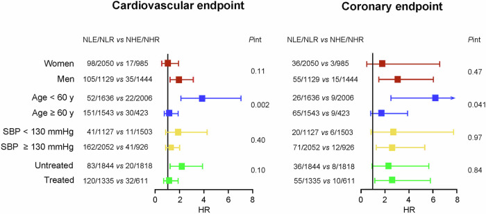
To investigate the consistency of the 1.3 PPA threshold, subgroup analyses were conducted in participants stratified by sex, age (<60 vs ≥60 years), median systolic BP (<130 vs ≥130 mmHg) and antihypertensive treatment status (Fig. 3). With full adjustments applied, the subgroup-by-PPA interaction terms were only significant if participants were categorized by age (P ≤ 0.041). The HRs for a cardiovascular endpoint in the age groups <60 vs ≥60 years were 3.86 (CI: 2.11–7.04) vs 1.19 (CI: 0.75–1.89); the corresponding HRs for the coronary endpoint were 6.21 (CI: 2.50–15.4) vs 1.77 (CI: 0.80–3.89). Among 2006 young (<60 years) low-risk individuals (PPA ≥ 1.3), 815 (40.6%) were women and 129 (6.43%) had isolated systolic hypertension (systolic/diastolic BP: ≥140/<90 mmHg, irrespective of treatment status), while among high-risk (PPA < 1.3) individuals in the same age band these numbers were 1107 (67.7%) and 208 (12.7%), respectively (P < 0.001 for both). Among 423 older (≥60 years) low-risk (PPA ≥ 1.3) participants, 172 (40.2%) were women and 128 (30.3%) had isolated systolic hypertension, while among high-risk (PPA < 1.3) individuals of similar age these numbers were 943 (61.1%) and 612 (39.7%), respectively (P < 0.001 for both). The proportion of women at high risk (PPA < 1.3) was higher in young individuals (<60 vs ≥60 years: 67.7% vs 61.5%; P < 0.001).
Fig. 3.
Hazard ratios expressing the risk of cardiovascular and coronary endpoints per the 1.3 pulse pressure amplification threshold by subgroups. Hazard ratios, given with 95% confidence interval accounted for cohort (random effect), sex, age, body mass index, heart rate, smoking and drinking, the total-to-HDL serum cholesterol ratio, the estimated glomerular filtration rate, antihypertensive drug intake, history of cardiovascular disease and diabetes. Adjustment for sex, age and antihypertensive treatment was omitted, if these variables defined the strata. Squares represent the point estimates of the hazard ratios (HR). Horizontal lines denote 95% confidence interval. NLE/NLR vs NHE/NHR refer to the number of participants with an event/number of participants at risk dichotomized by the threshold of pulse pressure amplification (<1.3 vs ≥1.3, respectively)
In multivariable models additionally adjusted for diastolic BP, the HR for coronary endpoints per threshold in the discovery dataset (PPA < 1.3 vs ≥1.3; number endpoints/individuals at risk: 66/2230 vs 12/1715) was 2.44 (CI: 1.18–5.07; P = 0.017) and 2.79 (CI: 1.10–7.06; P = 0.034) in the replication dataset (25/949 vs 6/714). The corresponding standardized HRs for PPA as continuously distributed variable were 0.57 (CI: 0.35–0.92; P = 0.022) and 0.64 (CI: 0.34–1.20; P = 0.12).
Discussion
According to our reading of the literature, the current study breaks new grounds in two ways. First, it is a prospective population study showing that lower PPA is a risk factor for fatal combined with nonfatal cardiovascular and coronary endpoints. Foremost, it defined and replicated an outcome-driven PPA threshold of 1.3, which particularly below age 60 indicated risk of a cardiovascular or coronary endpoint.
To our knowledge, only three longitudinal population studies reported on the risk associated with PPA. The PARTAGE (Predictive Values of Blood Pressure and Arterial Stiffness in Institutionalized Very Aged Population) Study included 1126 participants (77.6% women), who were living in French and Italian nursing homes (mean age: 88.5 years) [3]. During the 2-year follow-up, 247 subjects died, and 228 experienced a major cardiovascular endpoint. A 10% PPA increase was associated with decreases in the event rates amounting to 24% and 17% for total mortality and cardiovascular events, respectively. In the current study, participants with a risk-carrying PPA had a significant higher rate of isolated systolic hypertension (25.8% vs 10.6%) and the HRs contrasting the risk of cardiovascular and coronary endpoints per PPA threshold in individuals <60 vs ≥60 years were 3.86 vs 1.19 and 6.21 vs 1.77, respectively. In the Framingham Heart Study [19], involving 2232 participants (58% women; mean age: 63 years), 151 cardiovascular endpoints occurred over 7.8 years of follow-up. Several indexes reflecting aortic stiffness were predictive, but the standardized multivariable-adjusted hazard ratio for PPA was 0.86 (CI: 0.19–3.82; P = 0.84). The Anglo-Cardiff Collaborative Study involved healthy people and patients with risk factors, such as hypertension, hypercholesterolemia, smoking or diabetes and patients with a history of cardiovascular disease [20]. The 10,613 participants were from 18 to 101 years old (50.8% women). In this report, PPA was expressed as the reciprocal of the traditionally used definition, so that higher values represented individuals with a relatively higher aortic pressure for a given brachial pressure. The aorta-to-brachial PP ratio averaged 0.72 in healthy subjects (1.39 according to the current PPA definition) and was consistently and significantly higher in all other groups (range of means: 0.77–0.80 [1.25–1.30 according to the current definition]) except in smokers (mean value: 0.66 [1.51]).
Pulse pressure amplification and aging
There is large consistency among reports describing the age dependency of PPA. In both women and men, central systolic BP increased more with age than brachial systolic BP (P < 0.001) [21, 22]. The cross-sectional ACCT [7], including 4001 healthy, normotensive individuals, aged 18–90 years, showed that PPA decreased from 1.72 in teenagers to 1.25 in octogenarians. Another study combined volunteers recruited from the community and patients attending an open access clinic for the assessment of cardiovascular risk [23]. Patients with cardiovascular disease, diabetes or on drug treatment were excluded, but not those with hypertension. PPA decreased linearly with age (r = −0.70; P < 0.001) [23]. However, the association with age of the ratio of brachial PP to the non-augmented central PP was not statistically significant (r = 0.10; P = 0.10) [23]. In the current study, the correlation coefficient between PPA and age was −0.50 (P < 0.001, Supplementary Fig. S2).
Clinical relevance
That PPA declines with aging is well established [1–5]. However, among 3642 IDCARS participants younger than 60 years, 1636 (44.9%) had a risk-carrying PPA of <1.3, indicative of early vascular aging and stiffening of the central elastic arteries. Of these high-risk individuals, 1107 (67.7%) were women. Identifications of these individuals at risk is clinically highly relevant, so that cardiovascular risk factors can be timely managed to prevention cardiovascular and coronary complications.
Study limitations
Whilst the IDCARS database is a powerful resource, our study must be interpreted within the context of its potential limitations. First, the reconstruction of the aortic pulse wave from the radial pulse wave using the SphygmoCor technology requires the application of a generalized transfer function, which has been validated [24], but which has also been criticized [25]. For example, the SPC-301 micromanometer interfaced with the SphygmoCor device uses a single pressure sensor for applanation tonometry, but some validation studies of the generalized transfer function have utilized a servo-controlled automated tonometric system based on an arrayed sensor to avoid issues related to a manually operated single sensor [26, 27]. Second, the demographic characteristics, the period of recruitment, and the assessment of endpoint data differed between cohorts. However, the present analyses were adjusted for cohort as a random effect. By design participant-level meta-analyses allow applying the same statistical methods to all contributing cohorts, which is a strong point compared with meta-analyses of summary statistics [28]. Finally, over a median follow-up only 109 coronary endpoints occurred, probably because the low event rate among Chinese (0.49%) vs Europeans (2.64%) and South Americans (2.64%).
Conclusions
Over and beyond common risk factors, low PPA defined as a brachial-to-central PP ratio of <1.3 is a forerunner of cardiovascular and coronary complications and is an underestimated risk factor in women aged from 30 to 60 years. Our study therefore adds to the growing evidence supporting pulse wave analysis for risk stratification in clinical centers where the technology is readily available. If the technology is unavailable, such as for instance in low- and middle-income countries, isolated systolic hypertension can be used as a proxy for PPA [29], in particular in middle-aged and older individuals [30]. However, an important caveat pertains to adolescents and young adults with isolated systolic hypertension. The risk of a cardiovascular events in such patients is similar to that of age-matched individuals without isolated systolic hypertension [31] and lower than those with combined systolic-diastolic hypertension or isolated diastolic hypertension [32, 33]. Indeed, young adults with isolated systolic hypertension appear to comprise a heterogeneous patients group, including those with increased stroke volume, normal central blood pressure but exaggerated blood pressure amplification from the central to the peripheral arteries, and those with accelerated vascular aging and premature arterial stiffness [34]. In these young patients, assessing arterial stiffness is even more indicated [35] as well as out-of-office blood pressure monitoring to exclude white-coat hypertension [36].
Supplementary information
Acknowledgements
The authors gratefully acknowledge the expert clerical assistance of Mrs. Renilde Wolfs (Studies Coordinating Centre, Leuven, Belgium).
The International Database of Central Arterial Properties for Risk Stratification (IDCARS) Investigators
Lucas S. Aparicio14, Jessica Barochiner14, Blerim Mujaj15, Lutgarde Thijs15, Jan A. Staessen15, Fang-Fei Wei15, Wen-Yi Yang15, Zhen-Yu Zhang15, De-Wei An16, Yi-Bang Cheng16, Qian-Hui Guo16, Jian-Feng Huang16, Qi-Fang Huang16, Yuan-Yuan Kang16, Yan Li16, Chang-Yuan Liu16, Chang-Sheng Sheng16, Ji-Guang Wang16, Ying Wang16, Dong-Yan Zhang16, Wei Zhang16, Jan Filipovský17, Jitka Seidlerová17, Eeva P. Juhanoja18, Antti M. Jula18, Annika S. Lindroos18, Teemu J. Niiranen18, Sam S. Sivén18, Edoardo Casiglia19, Alessandra Pizziol19, Valérie Tikhonoff19, Babangida S. Chori20, Benjamin Danladi20, Augustine N. Odili20, Henry Oshaju20, Wiesława Kucharska21, Katarzyna Kunicka21, Natasza Gilis-Malinowska21, Krzysztof Narkiewicz21, Wojciech Sakiewicz21, Ewa Swierblewska21, Kalina Kawecka-Jaszcz22, Katarzyna Stolarz-Skrzypek22, Catharina M. C. Mels23, Ruan Kruger23, Gontse G. Mokwatsi23, Aletta E. Schutte23, Gavin R. Norton24, Angela Woodiwiss24, Daniel Ackermann25, Murielle Bochud25, Georg Ehret25, Ramón Álvarez-Vaz26, Anna C. Rios26, Florencia Carusso26, Mariana Sottolano26, José Boggia26, Luciana Borgarello26, Sebastián Robaina26, Paula Moliterno26, Oscar Noboa26, Alicia Olascoaga26, Alicia da Rosa26, Nadia Krul26, Matias Pécora26
Funding
Argentina: The Internal Medicine Service, Hospital Italiano de Buenos Aires, Buenos Aires, Argentina; Belgium: European Union (HEALTH-F7-305507 HOMAGE), European Research Council (Advanced Researcher Grant 2011-294713-EPLORE and Proof-of-Concept Grant 713601-uPROPHET), European Research Area Net for Cardiovascular Diseases (JTC2017-046-PROACT) and Research Foundation Flanders, Ministry of the Flemish Community, Brussels, Belgium (G.0881.13); Belgium (APPREMED): The Research Institute Alliance for the Promotion of Preventive Medicine (URL: www.appremed.org) received a non-binding grant from OMRON Healthcare Co., Ltd., Kyoto, Japan; China: The National Natural Science Foundation of China (Grants 82270469, 82070432, 81970353), the Ministry of Science and Technology (2022YFC3601302), Beijing, China, and by the Shanghai Commissions of Science and Technology (Grants 19ZR1443300), and the Shanghai Municipal Health Commission (202340035, 20234Y0036, 201940297 and a Grant for Leading Academics 2022LJ022); Czech Republic: European Union (Grants LSHM-CT-2006–037093 and HEALTH-F4-2007–201550) and Charles University Research Fund (Project P36); Finland: Academy of Finland (Grant 321351), Emil Aaltonen Foundation, the Paavo Nurmi Foundation, the Urmas Pekkala Foundation, and the Hospital District of South-Western Finland; Italy: European Union (Grants LSHM-CT-2006–037093 and HEALTH-F4-2007–201550); Poland (Gdańsk): European Union (Grants LSHM-CT-2006–037093 and HEALTH-F4-2007–201550); Poland (Kraków): European Union (Grants LSHM-CT-2006–037093 and HEALTH-F4-2007–201550) and Foundation for Polish Science; Uruguay: Asociación Española Primera en Salud. Switzerland: Swiss National Science Foundation (FN 33CM30‐124087 and FN 33CM30‐140331). The funding source had no role in the design and conduct of the study; collection, management, analysis, and interpretation of the data; preparation, review, or approval of the manuscript; and decision to submit the manuscript for publication.
Compliance with ethical standards
Conflict of interest
The authors declare no competing interests.
Footnotes
Members of the International Database of Central Arterial Properties for Risk Stratification (IDCARS) Investigators are listed below Acknowledgements.
Publisher’s note Springer Nature remains neutral with regard to jurisdictional claims in published maps and institutional affiliations.
These authors jointly supervised this work: Yan Li, Jan A. Staessen.
Contributor Information
Yan Li, Email: liyanshcn@163.com.
Jan A. Staessen, Email: jan.staessen@appremed.org
The International Database of Central Arterial Properties for Risk Stratification (IDCARS) Investigators:
Lucas S. Aparicio, Jessica Barochiner, Blerim Mujaj, Lutgarde Thijs, Jan A. Staessen, Fang-Fei Wei, Wen-Yi Yang, Zhen-Yu Zhang, De-Wei An, Yi-Bang Cheng, Qian-Hui Guo, Jian-Feng Huang, Qi-Fang Huang, Yuan-Yuan Kang, Yan Li, Chang-Yuan Liu, Chang-Sheng Sheng, Ji-Guang Wang, Ying Wang, Dong-Yan Zhang, Wei Zhang, Jan Filipovský, Jitka Seidlerová, Eeva P. Juhanoja, Antti M. Jula, Annika S. Lindroos, Teemu J. Niiranen, Sam S. Sivén, Edoardo Casiglia, Alessandra Pizziol, Valérie Tikhonoff, Babangida S. Chori, Benjamin Danladi, Augustine N. Odili, Henry Oshaju, Wiesława Kucharska, Katarzyna Kunicka, Natasza Gilis-Malinowska, Krzysztof Narkiewicz, Wojciech Sakiewicz, Ewa Swierblewska, Kalina Kawecka-Jaszcz, Katarzyna Stolarz-Skrzypek, Catharina M. C. Mels, Ruan Kruger, Gontse G. Mokwatsi, Aletta E. Schutte, Gavin R. Norton, Angela Woodiwiss, Daniel Ackermann, Murielle Bochud, Georg Ehret, Ramón Álvarez-Vaz, Anna C. Rios, Florencia Carusso, Mariana Sottolano, José Boggia, Luciana Borgarello, Sebastián Robaina, Paula Moliterno, Oscar Noboa, Alicia Olascoaga, Alicia da Rosa, Nadia Krul, and Matias Pécora
Supplementary information
The online version contains supplementary material available at 10.1038/s41440-024-01779-4.
References
- 1.Avolio AP, Van Bortel LM, Boutouyrie P, Cockcroft JR, McEniery CM, Protogerou AD, et al. Role of pulse pressure amplification in arterial hypertension: experts’ opinion and review of the data. Hypertension. 2009;54:375–83. 10.1161/HYPERTENSIONAHA.109.134379 [DOI] [PubMed] [Google Scholar]
- 2.Benetos A, Thomas F, Joly L, Blacher J, Pannier B, Labat C, et al. Pulse pressure amplification: a mechanical biomarker of cardiovascular risk. J Am Coll Cardiol. 2010;55:1032–37. 10.1016/j.jacc.2009.09.061 [DOI] [PubMed] [Google Scholar]
- 3.Benetos A, Gautier S, Labat C, Salvi P, Valbusa F, Marino F, et al. Mortality and cardiovascular events are best predicted by low central/peripheral pulse pressure amplification but not by high blood pressure levels in elderly nursing home subjects: the PARTAGE (Predictive Values of Blood Pressure and Arterial Stiffness in Institutionalized Very Aged Population) study. J Am Coll Cardiol. 2012;60:1503–11. 10.1016/j.jacc.2012.04.055 [DOI] [PubMed] [Google Scholar]
- 4.McEniery CM, Cockcroft JR, Roman MJ, Franklin SS, Wilkinson IB. Central blood pressure: current evidence and clinical importance. Eur Heart J. 2014;35:1719–25. 10.1093/eurheartj/eht565 [DOI] [PMC free article] [PubMed] [Google Scholar]
- 5.Chirinos JA, Segers P, Hughes T, Townsend R. Large-artery stiffness in health and disease: JACC state-of-the-art review. J Am Coll Cardiol. 2019;74:1237–63. 10.1016/j.jacc.2019.07.012 [DOI] [PMC free article] [PubMed] [Google Scholar]
- 6.Chirinos JA, Segers P. Noninvasive evaluation of left ventricular afterload: part 1: pressure and flow measurements and basic principles of wave conduction and reflection. Hypertension. 2010;56:555–62. 10.1161/HYPERTENSIONAHA.110.157321 [DOI] [PubMed] [Google Scholar]
- 7.McEniery CM, Yasmin Hall IR, Qasem A, Wilkinson IB, Cockcroft JR, on behalf of the ACCT Investigators. Normal vascular aging: differential effects on wave reflection and aortic pulse wave velocity: the Anglo-Cardiff Collaborative Trial (ACCT). J Am Coll Cardiol. 2005;46:1753–60. 10.1016/j.jacc.2005.07.037 [DOI] [PubMed] [Google Scholar]
- 8.Wassertheurer S, Burkhardt K, Heemann U, Baumann M. Aortic to brachial pulse pressure amplification as functional marker and predictor of renal function loss in chronic kidney disease. J Clin Hypertens. 2014;16:401–5. 10.1111/jch.12316 [DOI] [PMC free article] [PubMed] [Google Scholar]
- 9.Cho SW, Kim BK, Kim JH, Byun YS, Goh CW, Rhee KJ, et al. Non-invasively measured aortic wave reflection and pulse pressure amplification are related to the severity of coronary artery disease. J Cardiol. 2013;62:131–37. 10.1016/j.jjcc.2013.03.014 [DOI] [PubMed] [Google Scholar]
- 10.Aparicio LS, Huang QF, Melgarejo JD, Wei DM, Thijs L, We FF, et al. The International Database of Central Arterial Properties for Risk Stratification: research objectives and baseline characteristics of participants. Am J Hypertens. 2021;35:54–64. 10.1093/ajh/hpab139 [DOI] [PMC free article] [PubMed] [Google Scholar]
- 11.World Medical Association. World Medical Association Declaration of Helsinki: ethical principles for medical research involving human subjects. J Am Med Ass. 2013;310:2191–94. 10.1001/jama.2013.281053 [DOI] [PubMed] [Google Scholar]
- 12.Pauca AL, O’Rourke M, Kon ND. Prospective evaluation of a method for estimating ascending aortic pressure from the radial artery pressure waveform. Hypertension. 2001;38:932–37. 10.1161/hy1001.096106 [DOI] [PubMed] [Google Scholar]
- 13.Levey AS, Stevens LA, Schmid CH, Zhang Y, Castro AF III, Feldman HI, et al. A new equation to estimate glomerular filtration rate. Ann Intern Med. 2009;150:604–12. 10.7326/0003-4819-150-9-200905050-00006 [DOI] [PMC free article] [PubMed] [Google Scholar]
- 14.Schoenfeld DA. Sample-size formula for proportional-hazards regression model. Biometrics. 1983;39:499–503. 10.2307/2531021 [DOI] [PubMed] [Google Scholar]
- 15.Gu YM, Thijs L, Li Y, Asayama K, Boggia J, Hansen TW, et al. Outcome-driven thresholds for ambulatory pulse pressure in 9938 participants recruited from 11 populations. Hypertension. 2014;63:229–37. 10.1161/HYPERTENSIONAHA.113.02179 [DOI] [PMC free article] [PubMed] [Google Scholar]
- 16.Kikuya M, Hansen TW, Thijs L, Björklund-Bodegård K, Kuznetsova T, Ohkubo T, et al. Diagnostic thresholds for ambulatory blood pressure monitoring based on 10-year cardiovascular risk. Circulation. 2007;115:2145–52. 10.1161/CIRCULATIONAHA.106.662254 [DOI] [PubMed] [Google Scholar]
- 17.Pencina MJ, D’Agostino RB Sr, D’Agostino RB Jr, Vasan RS. Evaluating the added predictive ability of a new marker: from area under the ROC curve to reclassification and beyond. Stat Med. 2008;27:157–72. 10.1002/sim.2929 [DOI] [PubMed] [Google Scholar]
- 18.Hosmer DW Jr, Leleshow S. Applied logistic regression. New York, USA: Wiley; 1989. pp. 47–56.
- 19.Mitchell GF, Hwang SJ, Vasan RS, Larson MG, Pencina MJ, Hamburg NM, et al. Arterial stiffness and cardiovascular events: the Framingham Heart Study. Circulation. 2010;121:505–11. 10.1161/CIRCULATIONAHA.109.886655 [DOI] [PMC free article] [PubMed] [Google Scholar]
- 20.McEniery, Yasmin CM, McDonnell B, Munnery M, Wallace SM, Rowe CV, et al. Central pressure: variability and impact of cardiovascular risk factors: the Anglo-Cardiff Collaborative Trial II. Hypertension. 2008;51:1476–82. 10.1161/HYPERTENSIONAHA.107.105445 [DOI] [PubMed] [Google Scholar]
- 21.Li Y, Staessen JA, Sheng CS, Huang QF, O’Rourke M, Wang JG. Age dependency of peripheral and central systolic pressures: cross-sectional and longitudinal observations in a Chinese population. Hypertens Res. 2012;35:115–22. 10.1038/hr.2011.160 [DOI] [PubMed] [Google Scholar]
- 22.Wojciechowska W, Staessen JA, Nawrot T, Cwynar M, Sleidlerová J, Stolarz K, et al. Reference values in White Europeans for the arterial pulse wave recorded by means of the ShygmoCor device. Hypertens Res. 2006;29:475–83. 10.1291/hypres.29.475 [DOI] [PubMed] [Google Scholar]
- 23.Wilkinson IB, Franklin SS, Hall IR, Tyrrell S, Cockcroft JR. Pressure amplification explains why pulse pressure is unrelated to risk in young subjects. Hypertension. 2001;38:1461–66. 10.1161/hy1201.097723 [DOI] [PubMed] [Google Scholar]
- 24.O’Rourke MF, Kim M, Adji A, Nichols WW, Avolio A. Use of arterial transfer function for the derivation of aortic wave characrteristics. J Hypertens. 2004;22:431–2. 10.1097/00004872-200402000-00030 [DOI] [PubMed] [Google Scholar]
- 25.Segers P, Mahieu D, Kips J, Van Bortel LM. The use of a generalized transfer function: different processing, different results! J Hypertens. 2007;25:1783–87. 10.1097/HJH.0b013e3282ef5c5f [DOI] [PubMed] [Google Scholar]
- 26.Chen CH, Nevo E, Fetics B, Pak PH, Yin FC, Maughan WL, et al. Estimation of central aortic pressure waveform by mathematical transformation of radial tonometry pressure: validation of generalized transfer function. Circulation. 1997;95:1827–36. 10.1161/01.CIR.95.7.1827 [DOI] [PubMed] [Google Scholar]
- 27.Sharman JE, Lim R, Qasem AM, Coombes JS, Burgess MI, Franco J, et al. Validation of a generalized transfer function to noninvasively derived central blood pressure during exercise. Hypertension. 2006;47:1203–8. 10.1161/01.HYP.0000223013.60612.72 [DOI] [PubMed] [Google Scholar]
- 28.Riley RD, Lambert PC, Abo-Zaid G. Meta-analysis of individual participant data: rationale, conduct, and reporting. Br Med J. 2010;340:c221. 10.1136/bmj.c221 [DOI] [PubMed] [Google Scholar]
- 29.Odili AN, Chori BS, Danladi B, Yang WY, Zhang ZY, Thijs L, et al. Electrocardiographic left ventricular hypertrophy in relation to peripheral and central blood pressure indices in a Nigerian population. Blood Press. 2019;29:39–46. 10.1080/08037051.2019.1646610 [DOI] [PubMed] [Google Scholar]
- 30.Staessen J, Amery A, Fagard R. Editorial review. Isolated systolic hypertension in the elderly. J Hypertens. 1990;8:393–405. 10.1097/00004872-199005000-00001 [DOI] [PubMed] [Google Scholar]
- 31.Eeftinck Schattenkerk DW, van Gorp J, Vogt L, Peters RJG, van den Born BJH. Isolated systolic hypertension of the young and its association with central blood pressure in a large multi-ethnic population: the HELIUS study. Eur J Prev Cardiol. 2018;25:1351–59. 10.1177/2047487318777430 [DOI] [PMC free article] [PubMed] [Google Scholar]
- 32.Saladini F, Fania C, Mos L, Mazzer A, Casiglia E, Palatini P. Office pulse pressure is a predictor of favorable outcome in young- to middle-aged subjects with stage 1 hypertension. Hypertension. 2017;70:537–42. 10.1161/HYPERTENSIONAHA.117.09516 [DOI] [PubMed] [Google Scholar]
- 33.Yao Y, Stamler J, Garside DB, Daviglus ML, Franklin SS, Cernethon MR, et al. Isolated systolic hypertension in young and middle-aged adullts and 31-year risk for cardiovasvular mortality. J Am Coll Cardiol. 2015;65:327–35. 10.1016/j.jacc.2014.10.060 [DOI] [PMC free article] [PubMed] [Google Scholar]
- 34.McEniery, Yasmin CM, Wallace S, Maki-Petaya K, McDonnell B, Sharman JE, et al. Increased stroke volume and aortic stiffness contribute to isolated systolic hypertension in young adults. Hypertension. 2005;46:221–26. 10.1161/01.HYP.0000165310.84801.e0 [DOI] [PubMed] [Google Scholar]
- 35.Palatini P, Rosei EA, Avolio A, Bilo G, Casiglia E, Ghiadoni L, et al. Isolated systolic hypertension in the young: a position paper endorsed by the European Society of Hypertension. J Hypertens. 2018;36:1222–36. 10.1097/HJH.0000000000001726 [DOI] [PubMed] [Google Scholar]
- 36.Asayama K, Stolarz-Skrzypek K, Yang WY, Hansen TW, Brguljan-Hitij J, Odili AN, et al. What did we learn from the international databases on ambulatory and home blood pressure in relation to cardiovascular outcome? Hypertens Res. 2023;46:934–49. 10.1038/s41440-023-01191-4 [DOI] [PMC free article] [PubMed] [Google Scholar]
Associated Data
This section collects any data citations, data availability statements, or supplementary materials included in this article.


