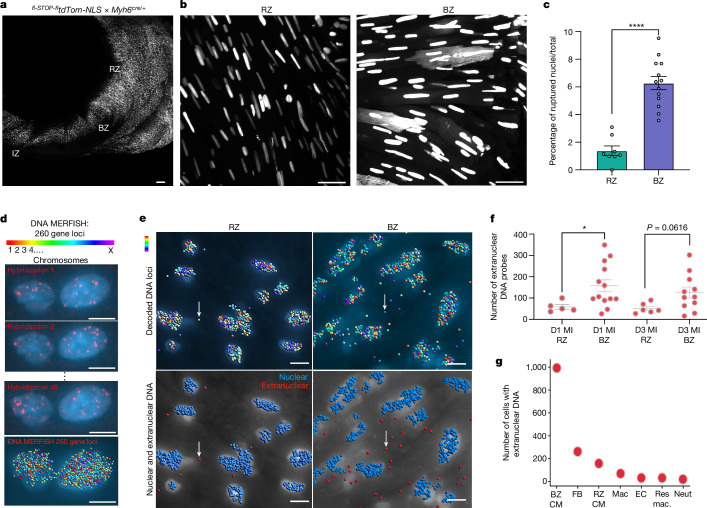
Fig. 4. Nuclear rupture and extranuclear DNA are found in load-bearing cells of the infarct BZ in vivo.
a, Representative image of short-axis whole-mounted heart from an infarcted cardiomyocyte-specific nuclear reporter mouse (CM-tdTom-NLS). This transgenic reporter was generated by crossing fl-STOP-fltdTom-NLSfl-STOP-fl and Myh6cre/+ mice. b, Representative images of CM-tdTom-NLS in nuclei of the RZ and BZ. Nuclear rupture was visualized as fluorescent reporter diffused throughout the cytoplasm of the outlined cardiomyocytes, whereas tdTom fluorescence was confined within the nuclear membrane in non-ruptured nuclei. c, Quantification of ruptured nuclei in the RZ versus BZ of infarcted CM-tdTom-NLS heart measured as the ratio between ruptured nuclei over total nuclei in each field of view. n = 788 RZ nuclei in 8 fields of view, 954 BZ nuclei in 13 fields of view, 1 mouse. d, Sequence-specific DNA probes were designed and synthesized to detect nuclear and extranuclear DNA in mouse hearts at day 1 and 3 after MI. Representative images of DNA MERFISH probes for 260 gene loci in 21 mouse chromosomes and rounds of hybridization of fluorescently labelled readout probes. e, Representative images of computationally decoded DNA loci (blue) in the RZ and BZ (e; top). Neighbour-based clustering of hybridized DNA probes was used to determine DNA probe localization to nuclear (blue) or extranuclear (red) compartments (e; bottom) and to quantify the number of extranuclear probes in imaged RZ and BZ regions (f) on days 1 and 3 after MI. n = 1,535 cells, 2 mice. g, Data of cells containing extranuclear DNA were ingested with RNA MERFISH data to determine the relative amounts of extranuclear DNA probes within each cell type. Data were analysed using unpaired two-tailed Mann–Whitney tests (c and f). Data are mean ± s.e.m. Results in a–c are representative of 70 observations of ruptured nuclei within the whole-mount section. Scale bars, 150 μm (a) and 10 μm (b, d and e).

