Fig. 1.
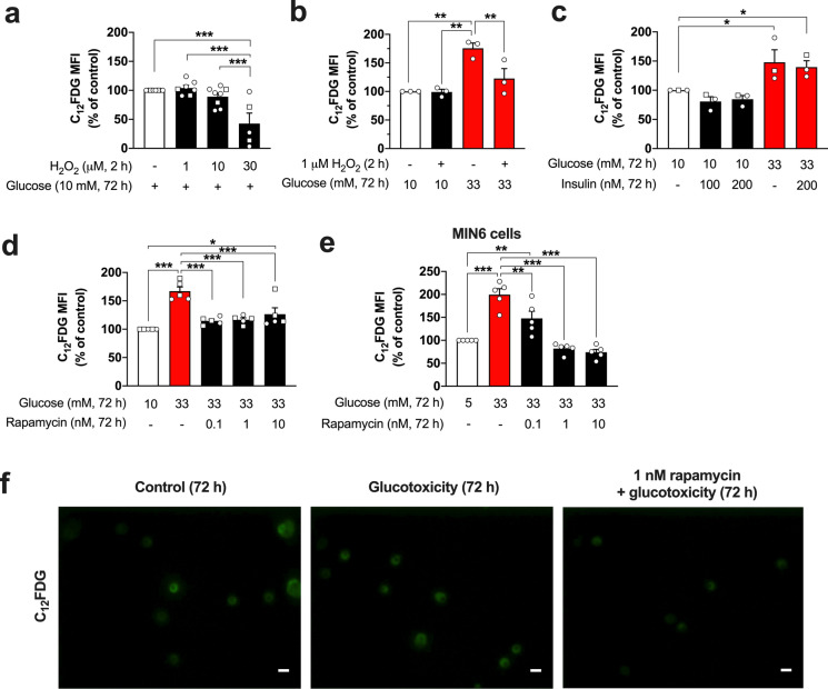
Influence of glucotoxicity and mTOR inhibition on the SA-beta-gal activity of primary islet cells. After a 2 h-exposure to hydrogen peroxide, the mouse islet cells were cultured for additional 72 h in standard culture medium (a). Alternatively, after the exposure to hydrogen peroxide (1 µM, 2 h) or vehicle, the islet cells were cultured for 72 h in a standard or glucotoxic (33 mM glucose) condition (b). Hydrogen peroxide does not increase SA-beta-gal but even reduces it at a concentration of 30 µM (a). In contrast, 33 mM glucose induces a significant rise in the activity of this senescence marker (b). The effects of insulin on SA-beta-gal were evaluated in standard or glucotoxic conditions, without showing any effect (c). Inhibition of mTOR signaling by rapamycin (0.1, 1 and 10 nM, 72 h) prevents the effects of glucotoxicity in islet cells (d) and MIN6 cells (e). Representative images of SA-beta-gal staining by C12FDG of islet cells cultured for 72 h under control, glucotoxic, or glucotoxic condition in the presence of rapamycin (f). MFI: mean fluorescence intensity. The number of independent mouse preparations (~ 7 months of age, a − d) or experiments (e) is indicated by the symbols. In mouse preparations, circles and squares represent female and male mice, respectively. *p < 0.05; **p < 0.01; ***p < 0.001. Scale bar (f) = 10 µm

