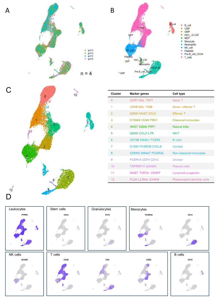
Fig. 4.
Identification of cell types in fresh L-PRF membranes (n = 4). (A) Cells are color coded by original sample identity. (B) Cell identification based on a supervised annotation algorithm (SingleR): cells are color coded by predicted cell type. (C) Clusters identified by an unsupervised graph-based algorithm: color coded by cluster (left), with cluster identification based on highly differentially expressed genes (right). (D) Feature plots indicating main leukocyte types. Pan leukocyte marker PTPRC (CD45); stem cell marker CD34; granulocyte marker FUT4 (CD15); monocyte markers FCGR3A and CD14; NK cell marker NCAM; T cell markers CD3E, CD4 and CD8A; B cell marker CD19

