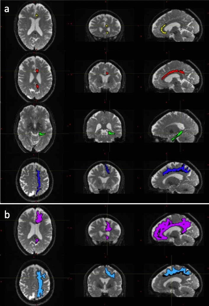
Fig. 2.

Tract Segmentations of the cingulate bundle and superior longitudinal fasciculus. Diffusion weighted image data for a 53-year-old female participant with left hemisphere tract segmentation mask overlays. a Displays the cingulum, divided into the peri-genual (CBG, yellow), dorsal (CBD, red) and temporal (CBT, green) tracts and the superior longitudinal fasciculus I (SLF-I, dark blue), extracted according to the Xtract definition. b Displays the cingulum bundle (CG, pink) and the superior longitudinal fascicules (SLF-I, light blue) extracted according to the Tract Seg definition
