Fig. 3.
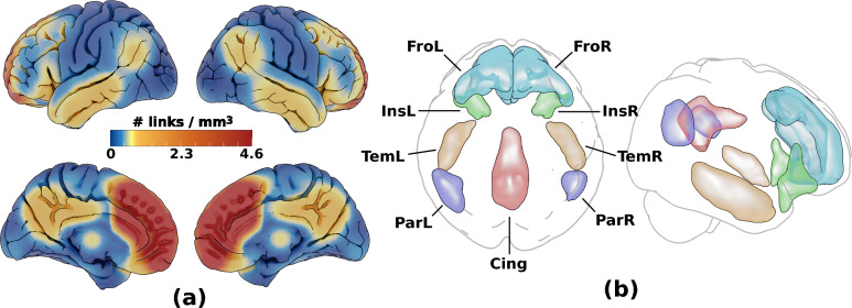
Voxel-based connectivity results. Cortical ROI set with high connectivity to the medial frontal lobe ROI, (visualised in Supplementary Fig. 2). a shows the link density from the medial frontal lobe ROI used to manually delineated highly connected regions in b: frontal (Fro: overlaps cing ant/mid, front sup med, front sup/mid, front mid/med/inf/sup orb), insular (Ins: overlaps insula, temp pole mid/sup, front inf orb/tri), temporal (Tem: overlaps temp inf/mid, temp pole mid), parietal (Par: overlaps par inf, temp mid/sup, angular, supramarginal) and posterior cingulate (Cing: overlaps cing mid/post, precuneus). To these, two subcortical ROIs defined in the Harvard–Oxford subcortical atlas were added (not shown): basal ganglia (BG: thalamus, Caudate, Pallidum, Putamen) and hippocampus/amygdala (HiAm)

