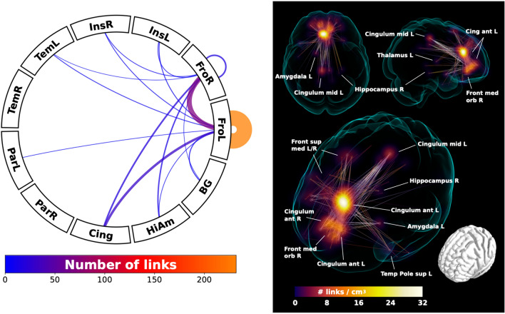
Fig. 4.
Voxel-based connectivity results. Network component with significantly greater functional connectivity in individuals with an absent left paracingulate sulcus relative to individuals with a present left paracingulate sulcus (P = 0.01 controlled for family-wise error rate). The left panel shows the connectogram in the form of the number of links connecting the left paracingulate ROI to highly connected regions (see Fig. 3). Number of links are represented by line thickness and colour, as denoted in the colour bar below. The right panel illustrates regions with the greatest link density (shown in voxel-wise maximal intensity projection) converging on the left anterior cingulate gyrus (Cingulum ant L), extending inferiorly towards the right frontal medial orbitofrontal (Front med orb R) and the right anterior cingulate gyrus (Cingulum ant R). More extended connections were also found to frontal superior medial gyrus L/R (Front sup med L/R) and the posterior cingulate gyrus (Cingulum mid L), as well as scattered connections to subcortical structures: left amygdala (Amygdala L), right posterior hippocampus (Hippocampus R), left thalamus (Thalamus L) and left superior temporal pole (Temporal Pole sup L). Lines are included to illustrate links between connected regions

