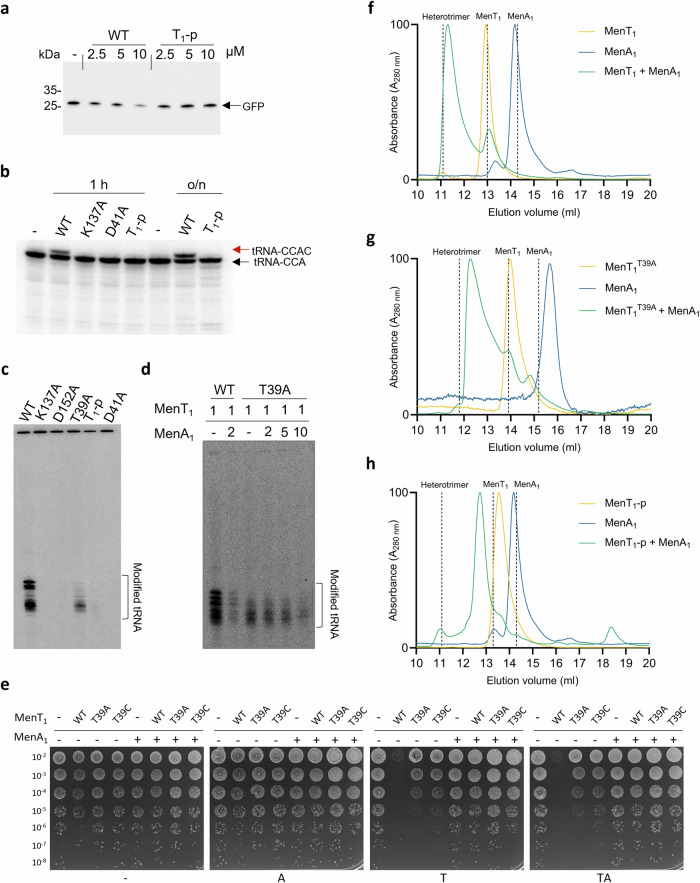
Fig. 5. Phosphorylation of MenT1 inhibits NTase activity.
a Total tRNA from E. coli was pre-incubated with MenT1 in vitro and subsequently used in a cell-free translation assay. Samples were separated on 4–20% SDS-PAGE gels and expression levels of GFP model substrate were determined using anti-GFP antibody. b [α-32P]-CTP labelled M. tuberculosis tRNA Gly-3 was incubated with MenT1 or its variants (5 µM) for 4 h at 37 °C in the presence of unlabelled CTP. Red arrows indicate the presence of cytidine extension. c, d Total RNA from M. smegmatis was incubated with MenT1 or its variants (5 µM) in the presence of [α−32P]-CTP at 37 °C for 2 h. e Toxicity/antitoxicity assays were performed in M. smegmatis to study the importance of MenT1 T39 in toxicity/antitoxicity. Co-transformants of M. smegmatis containing pGMC -vector (-), -MenT1 WT, or -MenT1 T39A and T39C variants, and pLAM -vector (-) or -MenA1 WT were serially diluted and spotted on LB agar plates in the presence or absence of toxin and antitoxin inducers (100 ng ml−1 ATc or 0.2% Ace, respectively). Plates were incubated for 3 days at 37 °C. “A” = antitoxin induced, “T” = toxin induced, “TA” = antitoxin + toxin induced. f–h Overlaid analytical SEC traces from a Superdex™ 75 increase 10/300 GL SEC column corresponding to either MenT1 (f), MenT1 T39A (g), or MenT1-p (h) incubated in the absence and presence of MenA1, confirming heterotrimeric complex formation blocks MenT1 T39A toxicity. Chromatograms are normalized between 0 and 100 for presentation and comparison, cropped to the appropriate scale. Vertical dashed lines display the expected elution volume of respective samples based on calculated Stokes Radii. Data are representative of three independent biological replicates. Source data are provided as a Source Data file.

