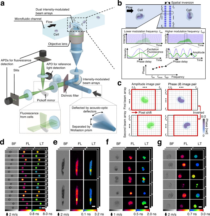
Fig. 1. High-throughput FLIM flow cytometry.
f1–fn denote modulation frequencies (n: the total number of modulation frequencies). a Schematic of the high-throughput FLIM flow cytometer. Dual intensity-modulated beam arrays enable double interrogation of a flowing cell, enhancing the precision of FLIM image acquisitions. APD: avalanche photodetector. b Principle of fluorescence lifetime pixel acquisition with a complementary modulation frequency pair (flow and fhigh). The time-varying phase delay of the fluorescence signal relative to the excitation was used for generating fluorescence lifetime images. fmean = (flow + fhigh)/2 = (f1 + fn)/2. c, Amplitude and phase image pairs generated from intensity-modulated beam arrays. t1–tm represent time points (m: the number of time points acquired for each triggered event). d–g Images of beads and cells acquired with the FLIM flow cytometer. BF bright-field, FL fluorescence, LT fluorescence lifetime. Scale bars: 10 μm. Two, two, four, and five independent experiments were performed, resulting in similar results for panels d–g, respectively. d Polymer beads with different fluorescence lifetimes. e Euglena gracilis cells stained with SYTO16. f Tumor-derived rat glioma cells stained with SYTO16. g Human cancer cells (Jurkat) stained with SYTO16.

