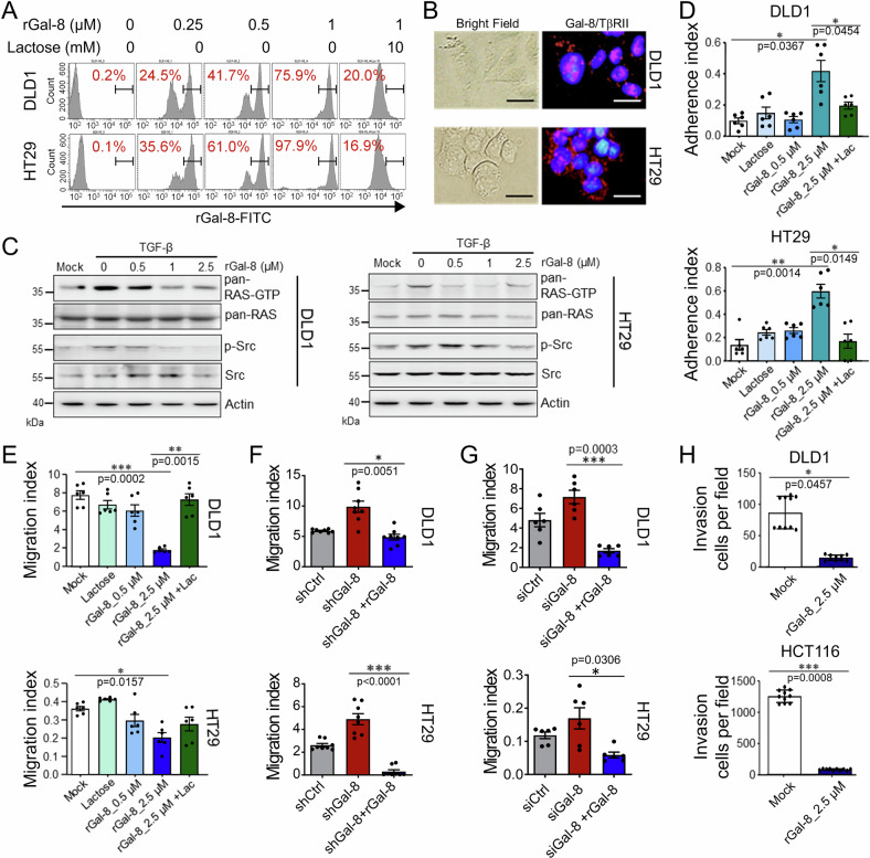
Fig. 4. rGal-8 suppresses migration and invasion of CRC cells by targeting TGF-β signaling.
A FACS shows binding of the indicated amounts of FITC-conjugated rGal-8 with DLD1 and HT29 cells. Lactose (10 mM) blocks the binding of rGal-8 with CRC cells. B PLA shows the interaction of galectin-8 (Gal-8) and TβRII on CRC cells. Scale bar = 10 μm. C Western blotting shows that rGal-8 affects TGF-β-mediated RAS and Src activities in CRC cells. Cell lysates from untreated DLD1 and HT29 cells (mock) or from cells treated with TGF-β (100 ng/mL) and various amounts of rGal-8 were subjected to immunoblotting with the indicated antibodies. Actin serves as the loading control. Cell adhesion (D) and migration (E) of DLD1 and HT29 cells treated with the indicated doses of rGal-8 and/or lactose (Lac, 100 mM), analyzed by xCELLigence RTCA. Bar graphs show the average cell indexes at 6 h (D) and 48 h (E). F Cell migration of DLD1 and HT29 cells expressing shCtrl or shGal-8 in the absence or presence of rGal-8 (2.5 µM) analyzed by xCELLigence RTCA at 48 h. G Cell migration of DLD1 and HT29 cells treated with siCtrl or siGal-8 in the absence or presence of rGal-8 (2.5 µM) analyzed by xCELLigence RTCA at 48 h. H Invasive activities of DLD1 and HCT116 cells treated with rGal-8 for 24 h analyzed by xCELLigence RTCA. Results in D, E, F, G, H are shown as mean ± SD (3, 3, 4 and 3 biological replicates with technical duplicates for each in D, E, F, G, respectively; 2 biological replicates with technical quintuplicates in H). Statistical tests were calculated by t-test.

