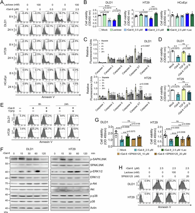
Fig. 5. rGal-8 triggers apoptosis in CRC cells by inducing transient JNK activation.
The level of cell apoptosis, determined by Annexin V staining (A), and cell viability, determined by MTT assay (B), of DLD1, HT29, and HCoEpi cells treated with various doses of rGal-8 in the presence or absence of lactose (Lac, 100 mM). C The activities of caspases in DLD1 and HT29 cells treated with rGal-8 (2.5 µM) or etoposide (10 µM, positive control of caspase activation) for the indicated time points were analyzed based on the cleavage of each substrate. D Cell viability of DLD1 and HT29 cells treated with rGal-8 (2.5 µM) or/and TGF-β (100 ng/mL) for 72 h determined by MTT assay. E FACS shows the percentage of Annexin V+ apoptotic DLD1 or HT29 cells treated with rGal-8 (2.5 µM) and/or TGF-β (100 ng/mL) for the indicated time points. F Western blotting shows the expression level of phosphorylated and total JNK, ERK1/2, AKT and p38 in DLD1 and HT29 cells treated with rGal-8 (2.5 µM) for the indicated time points. Cell viability determined by MTT assay (G), and apoptosis determined by Annexin V staining (H) of DLD1 and HT29 cells treated with rGal-8 (2.5 µM) alone or together with JNK inhibitor (SP600125) at the indicated doses for 72 h (G) and 24 h (H). Lactose (Lac, 100 mM) was used to block the effect of rGal-8 in some groups. Results in B, C, D, G are shown as mean ± SD (3 biological replicates with technical duplicates for each in B, C, G; two biological replicates with technical duplicates for D). Statistical tests were calculated by t-test.

