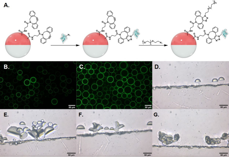
Figure 2.
A. Droplet bioconjugation scheme. PDBCO-PPEG-b-PBn-functionalized droplets were sequentially reacted with azide-functionalized rcSso7d proteins, followed by poly(ethylene glycol) methyl ether azide. Images are not drawn to scale; droplets are orders of magnitude larger than protein components. Fluorescence microscopy images of droplets reacted with B. FITC-labeled E1 and C. FITC-labeled E2. Side-view images of D. E1+PEG-functionalized Janus droplets, E. and F. 1:1 E1+PEG- and E2+PEG-functionalized droplets incubated with 5 μg/mL of IL-6, and G. DNA-functionalized droplets in 0.1 wt % of 4:6 CTAB:Zonyl that are agglutinated due to electrostatic interactions between the DNA and CTAB. All images are of polydisperse droplets.

