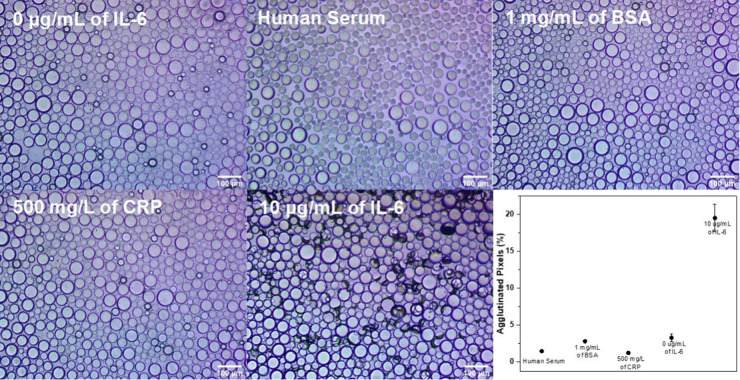
Figure 3.
Inverted microscope images of 1:1 E1+PEG- and E2+PEG-functionalized droplets incubated under different conditions overnight (0 μg/mL of IL-6, human serum, 1 mg/mL of BSA, 500 mg/L of CRP, and 10 μg/mL of IL-6). The agglutinated droplets appear on their side and create darkened spots in the images as a result of their efficient scattering of transmitted light. At the bottom right is a plot of the percent of agglutinated pixels according to image processing for each control relative to the 10 μg/mL of IL-6 assay.

