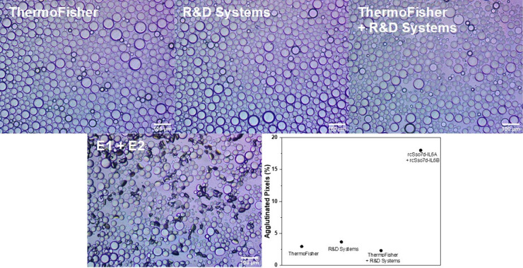
Figure 6.
Inverted microscope images of anti-IL-6 antibody+PEG-functionalized droplets and 1:1 E1+PEG- and E2+PEG-functionalized droplets incubated with 5 μg/mL of IL-6 overnight. Droplets with antibodies from ThermoFisher and R&D Systems and rcSso7d proteins purified via Ni2+ resin and subsequent FPLC are shown. Plot of the percent of agglutinated pixels according to image processing for each antibody-based assay relative to the 5 μg/mL of IL-6 assay.

