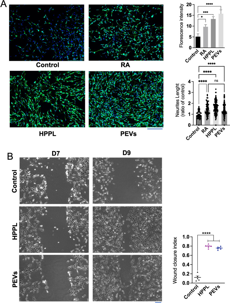
Fig. 5.
Functional activity of PEVs to promote neuronal growth in vitro on SH-SY5Y neuroblastoma cell line. A Capacity of PEVs to stimulate the neuronal maturation of SH-SY5Y cells. Cells were immuno-stained with β-III tubulin and counterstained with DAPI. Images showing extension of SH-SY5Y neurites under the treatment of PEVs and HPPL. HPPL and RA were used as positive controls to stimulate cell differentiation. Scale bar = 250 μm. Quantitative measurement of the fluorescence intensity (right) showed the capacity of PEVs to induce SH-SY5Y neuronal maturation. N = 3, *p < 0.05; ***p < 0.001 compared to the untreated negative control. From the captured images of β-III tubulin fluorescence, the length of each extension was measured individually to estimate total neurite outgrowth. The ratio of neurite length in treated cells to that in untreated cells was then calculated. N = 3, significant difference (ns), ****p < 0.001 compared to the untreated negative control. B Neuro-restoration effect of PEVs on the differentiated SH-5YSY. A scratch assay was performed using differentiated SH-SY5Y cells. Cells without any treatment (negative control), HPPL (positive control), and PEVs were used. The neuro-restoration effect was monitored by microscopy two days after the treatments on Day 9 (D9). Scale bar = 100 μm. The results are expressed as a wound-healing index, determined by the formula: (initial wound area − final wound area)/initial wound area). N = 3, ****p < 0.001 compared to untreated negative control. RA: Retinoic acid; PEVs: Platelet-extracellular vesicles; HPPL: heat-treated platelet pellet lysate

