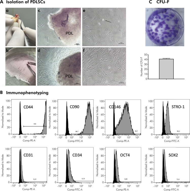
Figure 1. Isolation and characterization of PDLSCs. A) Representative images of a) Macroscopic aspect after scaling the middle third of the root of teeth collected to obtain PDLSCs; b) Fragments of periodontal ligament obtained from root scaling; c) Microscopic aspect of fragments of periodontal ligament obtained during establishment of the primary culture. Cells are observed migrating from the explant after 7 days of cell culture (scale bar = 100 μm); d) Microscopic aspect of the explant after 10 days of cell culture (scale bar = 100 μm); e) Microscopic aspect of PDLSCs after trypsinization, the detail shows cell mitosis (arrow) (scale bar = 100 μm); f) Microscopic aspect of PDLSCs in Passage 0, showing the spindle-like morphologic appearance of isolated PDLSCs (scale bar = 100 μm). B) Immunophenotyping. C) Macroscopic aspect of CFU-F and its quantification.
Analysis of the histograms performed in FlowJo software. Black curve represents the negative control (cells without fluorescent particles).

