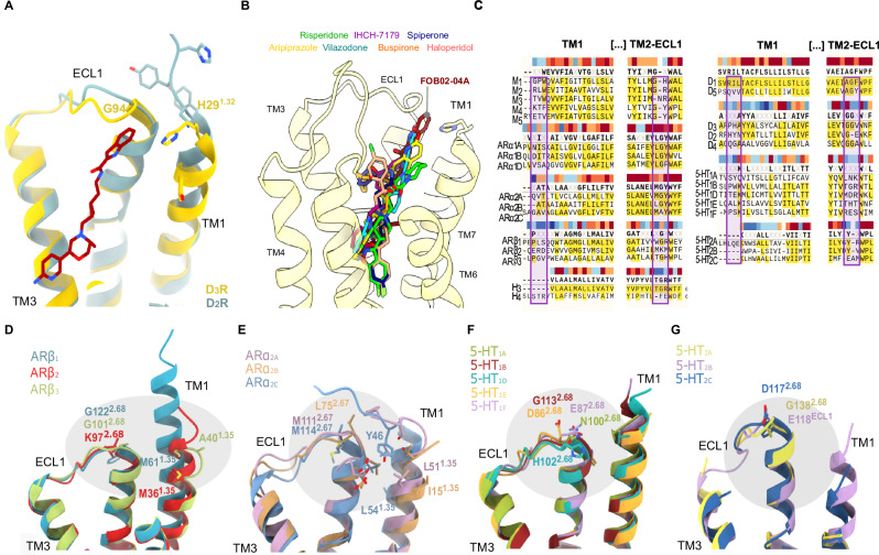
Fig. 4. Sequence and structural diversity of the SBP2-ECL1-1 in aminergic GPCRs.
A Comparison of the D3R (yellow cartoons with relevant residues as sticks) and D2R (light blue cartoons with relevant residues as sticks) TM2-ECL1 and TM1 regions within reach of FOB02-04A (dark red, sticks). B Relative binding sites of other bitopic ligands bound to D2R (haloperidol (PDB 6LUQ), spiperone (PDB 7DFP), risperidone (PDB 6CM4) and 5-HT1AR (aripiprazole (PDB 7E2Z), IHCH-7179 (PDB 8JT6), vilazodone (PDB 8FYL) and buspirone (PDB 8FYX) as shown on the D3R:FOB02-04A cryo-EM structure. C Sequence alignment of TM1 and TM2-ECL1 regions in aminergic GPCRs with residues around the SBP2-ECL1-1 embedded in a box. Sequence conservation is color-coded above each residue position (gradient from dark red, conserved, to dark blue, non-conserved). Structural differences at the SBP2-ECL1-1 site among closely related adrenergic receptors (D, E) and serotonin receptors (F, G). Receptors are shown as cartoons colored by receptors with relevant residues shown as sticks.

