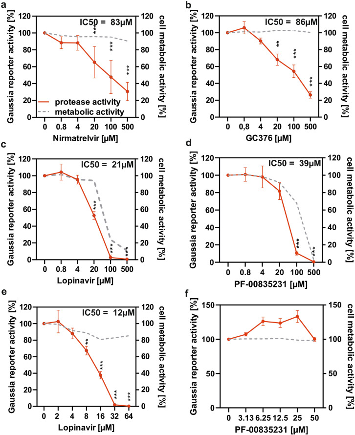
Fig. 4.
Evaluation of protease inhibitors using the Nsp5 reporter assay. HEK293T reporter cells were co-transfected with equal amounts of plasmid encoding the ACE2-Gal4 fusion protein and SARS-CoV-2 Nsp5 or GFP (negative control). After 6 h cells were treated with indicated molar concentration of inhibitors (a) nirmatrelvir, (b) GC376, (c) Lopinavir and (d) PF-00835231 or DMSO control for 20 h. The activity of secreted Gaussia luciferase was measured using luminometer and cellular metabolic activity was evaluated by the MTT assay. The activity of Lopinavir and PF-00835231 the was re-evaluated at lower, nontoxic concentrations (e and f). Red line indicates relative Nsp5 activity as percentage of no inhibitor control measured by luciferase assay (left y-axis) and grey dotted line indicates metabolic activity as percentage of inhibitor untreated cells as determined by MTT assay (right y-axis). Mean of 3–5 independent experiments + SD. *P < 0.05; **P < 0.01; ***P < 0.001, paired Student’s t-test. IC50 values were calculated using Prism GraphPad.

