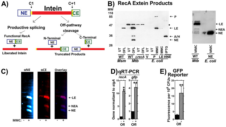
Fig. 1.
Validation of the splicing reporter system. (A) Schematic of RecA splicing products. Mtb RecA is translated as a precursor containing an internal 48 kDa RecA intein. The intein is flanked by an N-terminal extein (NE) of 26.5 kDa and a C-terminal extein (CE) of 11 kDa. Splicing depends on the first residues of the intein (cysteine, C1), and the C-extein (cysteine, C + 1). Productive splicing results in precise liberation of the intein from the precursor and allows the N- and C-terminal exteins to be ligated into functional RecA protein (ligated exteins, LE). Alternatively, off-pathway products result in liberation of either the C-extein (C-terminal cleavage) or of the N-extein (N-terminal cleavage). (B) Left: Western blot of whole cell lysates from M. smegmatis (Msm), Mtb H37Rv mc26230 (Mtb) or recombinant E. coli probed with anti- N-extein antibody. RecA splicing was evaluated in response to ofloxacin (OFL), Mitomycin C (MMC), or in untreated cultures (UT) to determine which bands contained splicing or aberrant cleavage products. Msm, which lacks the recA intein sequences, was used as a size marker for LE. ‘WT’ refers to presence of wild-type recA locus in Msm mc2155. E. coli expressing precursor (P), LE, or NE were included for additional reference bands, as marked, and to compare RecA intein splicing activity in a non-native bacterial host with that in Mtb. RecA was examined in Mtb with a wild-type recA locus (WT) or recA deletion (ΔrecA) to identify recA-specific bands. Black dot ● denotes a non-specific background band in Mtb cultures. A/H marks position of RecA-specific band of unknown identity (NEA) and a 6xHis-tagged N-extein protein (HNE) expressed in E. coli BL21 that migrated to similar positions in the gel. Right: E. coli expressing tagless version of NE was used to confirm Mtb NE band. Other abbreviations: Vehicle control (V), Isopropyl β-D-1-thiogalactopyranoside treatment (IPTG). (C) Lysates of MMC-treated Mtb cultures were dual-probed for the (left) NE and (center) CE using fluorescent secondary antibodies. Fluorescent images were overlayed (right) to identify bands cross-reacting bands. LE and NEA bands were visualized with both antibodies. (D) Expression of native recA and recA promoter-driven gfp in mc26230 cultures containing the PrecA:GFP reporter was examined via RT-PCR and (E) GFP fluorescence in response to ofloxacin. **indicates p < 0.01 Panel C: n = 2. Panel D: n = 3.

