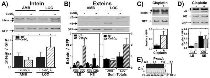
Fig. 3.
RecA splicing in response to copper and cisplatin. (A, B) Mtb cultures were grown shaking in ambient (AMB) or hypoxic (LOC) conditions in the presence ( +) or absence (−) of 50 µM copper sulfate (CuSO4) for 7 days. Top: Representative western of intein (A) or LE, and NE products in response to copper treatment in AMB or LOC (B). Bars represent mean and standard deviation. Bottom left: Densitometric quantification of LE and NE from 3 biological western blot replicates. Bottom right: Sum of LE and NE bands from the 3 replicates. Bars represent mean and standard error of the mean. (C, D) Mtb cultures were treated with cisplatin (CIS), and levels of excised intein (C) and LE and NE (D) were examined by western blot and normalized to GFP (top). Densitometric quantification of 3 biological western blot replicates. Bars represent mean and standard deviation (bottom). (E) recA expression in response to cisplatin as read out by our GFP transcriptional reporter assay. ● indicates background bands in the extein or intein western blot, n = 3. * indicates p < 0.05.

