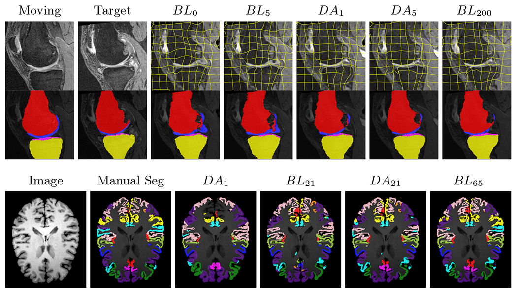
Fig. 2:

Examples of knee MRI registration (top) and brain MRI segmentation (bottom) results. Top: The first two columns are the moving image/segmentation and the target image/segmentation followed by the warped moving images (with deformation grids)/segmentations by different models. Bottom left to right: original image, manual segmentation, and predictions of various models. BLi and DAi represent the baseline and DA models with i manual segmentations respectively.
