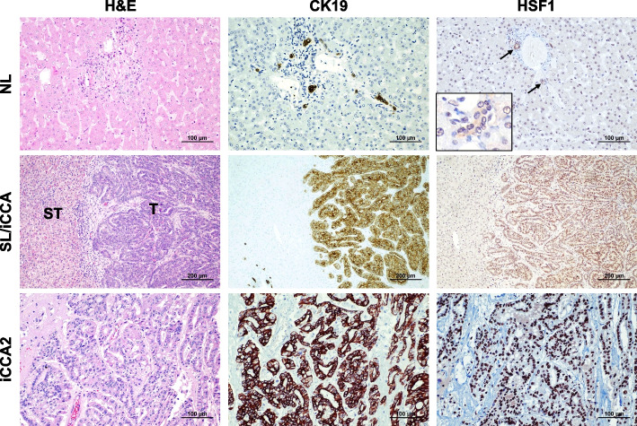
Fig. 2.
Representative immunohistochemistry patterns of HSF1 protein in human intrahepatic cholangiocarcinoma (iCCA; n = 186) and corresponding non-tumorous livers. The upper panels show a human normal liver (NL) displaying weak to moderate HSF1 immunoreactivity in hepatocytes and biliary cells (indicated by arrows). Staining of cholangiocytes (indicated by arrows) is better appreciable in the inset. Middle panels show the immunohistochemical staining of HSF1 in a non-tumorous surrounding tissue and one human iCCA specimen. The staining pattern for HSF1 is significantly more pronounced in the tumor compartment (T) compared with the neighboring non-tumorous surrounding tissue (ST), which exhibits faint HSF1 staining. Lower panels depict a second tumor (iCC2), characterized by intense nuclear HSF1 immunoreactivity. CK19 staining was used as a biliary marker. Abbreviation: H&E, hematoxylin and eosin staining. Original magnifications: 200x in the upper and lower panels, 100x in the middle panels, and 400x in the inset. Scale bar: 100 µm in the top and low panels, 200 µm in the middle panels

