Fig. 6.
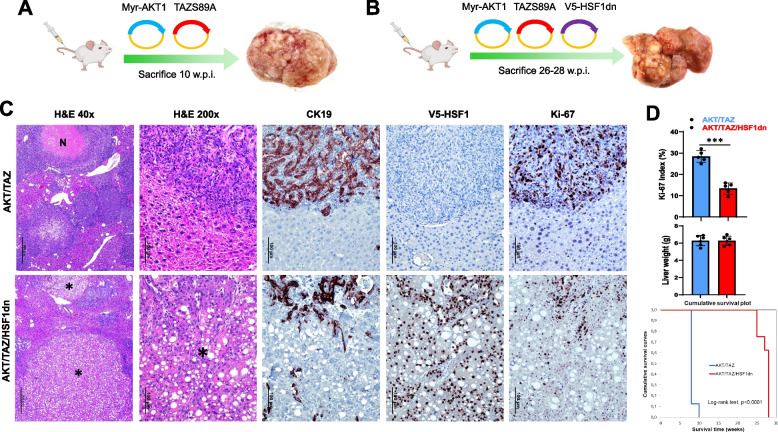
Suppression of HSF1 activity delays cholangiocarcinogenesis and reduces the proliferation and aggressiveness of AKT/TAZ mouse lesions. A, B Study design. Briefly, wild-type FVB/N mice were subjected to hydrodynamic tail vein injection of either AKT/TAZS89A/pT3 (control; AKT/TAZ mice) or AKT/TAZS89A/HSF1dn (AKT/TAZ/HSF1dn mice) plasmids. HSF1dn is the dominant-negative form of the HSF1 transcription factor, whose overexpression effectively inhibits the endogenous HSF1 activity. C Liver lesions from AKT/TAZ mice consisted of invasive and proliferative (as assessed by positive immunoreactivity for Ki-67) intrahepatic cholangiocarcinomas (upper panels) with frequent necrotic areas (N). In contrast, neoplastic lesions from AKT/TAZ/HSF1dn consisted of both hepatocellular, clear-cell (indicated by asterisks), and cholangiocellular lesions with low proliferation rates. The hepatocellular lesions were CK19-negative, whereas the cholangiocellular lesions were CK19-positive. As expected, V5-tagged-HSF1dn staining was observed only in AKT/TAZ/HSF1dn mice. D While liver weight was equivalent in the two mouse cohorts, the proliferative activity was significantly higher in AKT/TAZ mice than in AKT/TAZ/HSF1dn mice. Moreover, the survival curve showed significantly longer survival of AKT/TAZ/HSF1dn mice. Student’s t-test: **, P < 0.0001. Original magnifications: 40x and 200x; scale bar: 500 μm in 40x and 100 μm in 200x. Abbreviations: H&E, hematoxylin and eosin staining

