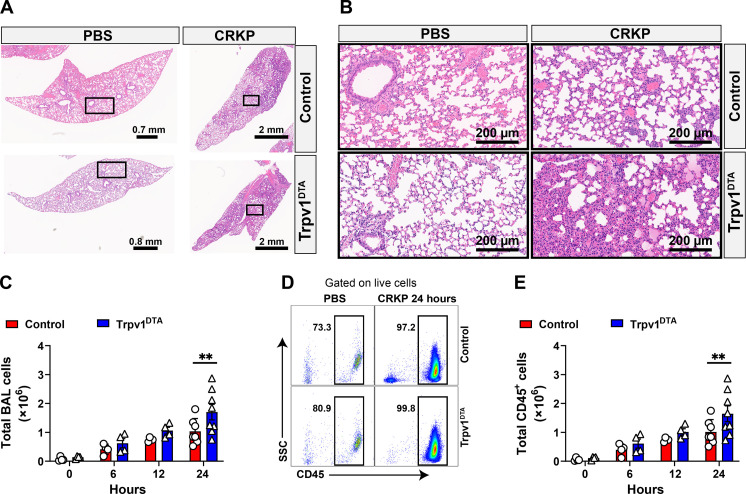
Fig. 2. Nociceptors suppress leukocyte influx during CRKP lung infection.
(A and B) H&E-stained sections of lungs: ×0.9 to ×1.1 magnification (A) and ×20 magnification of selected area (in rectangles) (B) of control and Trpv1DTA mice with PBS inoculation and after CRKP infection (24 hpi). Representative images were selected from at least 12 lobes of n = 3 to 4 mice in each group. Scale bars are shown in each figure. (C to E) Total live cells (C), representative flow cytometry plots of total leukocytes (CD45+ cells) (D), and total CD45+ cells (E), in BALF of Trpv1DTA and control mice at 6, 12, and 24 hpi with CRKP. Data in (C) and (E) are the means ± SEM and involve n = 4 mice per group for 0-, 6-, and 12-hour time course analyses and n = 7 to 8 mice per group for 24-hour time course analyses. Statistical analysis was done by two-way ANOVA of two-stage liner step-up procedure of Benjamini, Krieger, and Yekutieli posttests with the following significance levels: **P < 0.01. BAL, bronchoalveolar lavage.

