Figure 1.

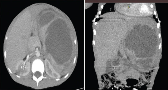
Contrast-enhanced abdominal CT scan (axial and coronal reconstructed images) at the level of the spleen showing multiloculated hypodense collection (HU: 7–29). CT: Computerised tomography

Contrast-enhanced abdominal CT scan (axial and coronal reconstructed images) at the level of the spleen showing multiloculated hypodense collection (HU: 7–29). CT: Computerised tomography