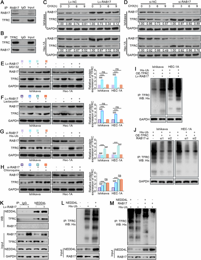
Fig. 4. RAB17 regulates TFRC expression in EC cells through the ubiquitin-proteasome pathway.
A Ishikawa cell lysates were incubated with an anti-RAF17 antibody, and interacting proteins were detected by Western blot analysis with an anti-TFRC antibody. B Ishikawa cell lysates were incubated with an anti-TFRC antibody, and interacting proteins were detected by Western blot analysis with an anti-RAB17 antibody. C Ishikawa and HEC-1A cells were infected with Lv-NC and Lv-RAB17. CHX (20 μmol) was added for the indicated time, and the cell lysates were subjected to Western blot analysis for RAB17 and TFRC. D Ishikawa and HEC-1A cells were transfected with NC-si or RAB17-si1. CHX (20 μmol) was added for the indicated time, and the cell lysates were subjected to Western blot analysis for RAB17 and TFRC. E Ishikawa and HEC-1A cells were infected with Lv-NC and Lv-RAB17. The cells were then treated with the MG132 proteasome inhibitor (20 mmol) for 12 h, and Western blot analysis was performed with anti-RAB17 and anti-TFRC antibodies. F Ishikawa and HEC-1A cells were transfected with Lv-NC or Lv-RAB17. The cells were then treated with the lactacystin proteasome inhibitor (10 mmol) for 12 h, and Western blot analysis was performed with anti-RAB17 and anti-TFRC antibodies. G Ishikawa and HEC-1A cells were transfected with NC-si or RAB17-si1. The cells were then transfected with His-tagged ubiquitin-containing vectors (His-Ub) for 12 h, and Western blot analysis was performed with anti-RAB17 and anti-TFRC antibodies. H Ishikawa and HEC-1A cells were transfected with NC-si or RAB17-si1. The cells were then treated with the chloroquine lysosomal inhibitor (10 mmol) for 12 h, and Western blot analysis was performed with anti-RAB17 and anti-TFRC antibodies. I, J Ishikawa and HEC-1A cells were transfected as indicated and treated with MG132 for 12 h. Lysates were immunoprecipitated with anti-TFRC and detected with anti-His. GAPDH was used as an internal control. K Ishikawa cells were infected with Lv-CTL or Lv-RAB17, and cell lysates were immunoprecipitated with the indicated primary antibody and immunoblotted as indicated. L, M Ishikawa cells were transfected as indicated, and then cell lysates were immunoprecipitated with anti-TFRC antibody and detected with anti-His antibody. All the above assays were independently performed in triplicate (N = 3). ***P < 0.001.

