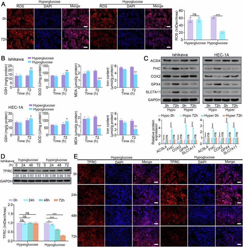
Fig. 5. RAB17 mediates TFRC-dependent ferroptosis in a hypoglycemic state.
A Representative images of immunofluorescence staining with an ROS probe in Ishikawa cell lines cultured in hyperglycemic (Hyper) or hypoglycemic (Hypo) medium for the designated times. Scale bars, 200 μm. B GSH, SOD, MDA, and levels in Ishikawa and HEC-1A cells cultured in hyperglycemic (Hyper) or hypoglycemic (Hypo) medium for the designated times. C Western blot analysis of designated marker proteins for ferroptosis in Ishikawa and HEC-1A cells cultured in hyperglycemic (Hyper) or hypoglycemic (Hypo) medium for the designated times. D Western blot analysis of TFRC expression in Ishikawa cells cultured in hyperglycemic (Hyper) or hypoglycemic (Hypo) medium for the designated times. E Representative images of immunofluorescence staining for TFRC in Ishikawa cell lines cultured in hyperglycemic (Hyper) or hypoglycemic (Hypo) medium for the designated times. Scale bars, 200 μm. GAPDH was used as an internal control. All the above assays were independently performed in triplicate (N = 3). The data are presented as the means ± SDs. The statistical analyses were performed by two-tailed unpaired Student’s t tests. *P < 0.05, **P < 0.01, and ***P < 0.001.

