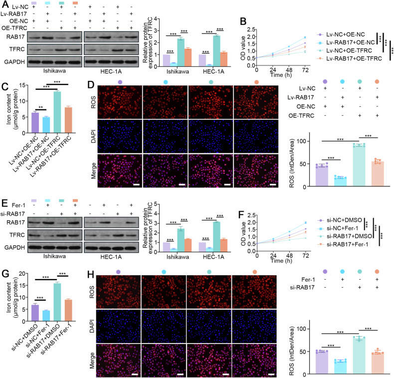
Fig. 6. TFRC-mediated ferroptosis is critical for RAB17-mediated regulation of EC cell proliferation.
A Western blot analysis of RAB17 and TFRC expression in Ishikawa and HEC-1A cells cotransfected with the designated vectors. B CCK-8 assays of Ishikawa cell lines cotransfected with the designated vectors. C The iron contents of Ishikawa cell lines cotransfected with the designated vectors. D Representative images of immunofluorescence staining with an ROS probe in Ishikawa cell lines cotransfected with the designated vectors. Scale bars, 200 μm. E Western blot analysis of RAB17 and TFRC expression in Ishikawa and HEC-1A cells transfected with/without designated siRNAs or treated with/without Fer-1. F CCK-8 assays of Ishikawa cell lines transfected with/without designated siRNAs or treated with/without Fer-1. G Iron content of Ishikawa cell lines transfected with/without designated siRNAs or treated with/without Fer-1. H Representative images of immunofluorescence staining using an ROS probe in Ishikawa cell lines transfected with/without the indicated siRNAs or treated with/without Fer-1. Scale bars, 200 μm. GAPDH was used as an internal control. All the above assays were independently performed in triplicate (N = 3). The data are presented as the means ± SDs. The statistical analyses were performed by two-tailed unpaired Student’s t tests. **P < 0.01, and ***P < 0.001.

