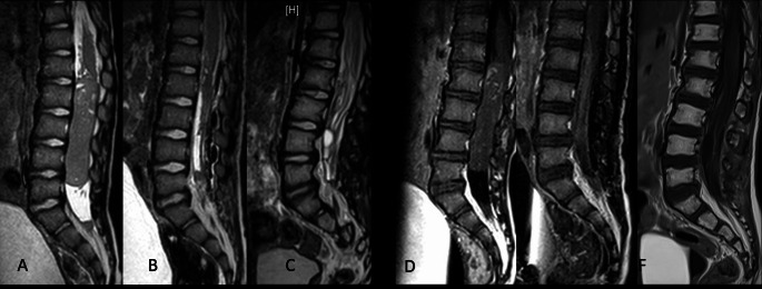
Fig. 2.
Preoperative (A, D), postoperative (B, E), and one-year follow-up (C, F) MRI images, both sagittal T2 and with contrast, show a spinal ETMR located in the lumbar spine of a 28-months-old child (ID_04). This child experienced severe pain, controllable only with morphine, and acute paraplegia. An urgent laminotomy from L1 to L5 and tumor resection were performed, resulting in total removal of the tumor. This was followed by multimodal adjuvant therapy

