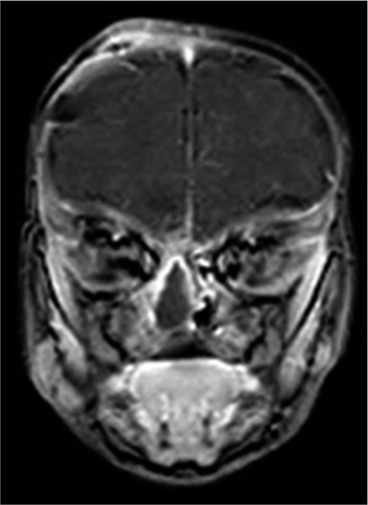
Figure 2:

Postoperative magnetic resonance imaging of the brain with coronal images done after the transcranial surgery showing a layered obliteration and separation of the anterior cranial fossa floor from the nasal cavity beneath. The remnant (to be removed later) soft-tissue sac can still be appreciated in the nasal cavity.
