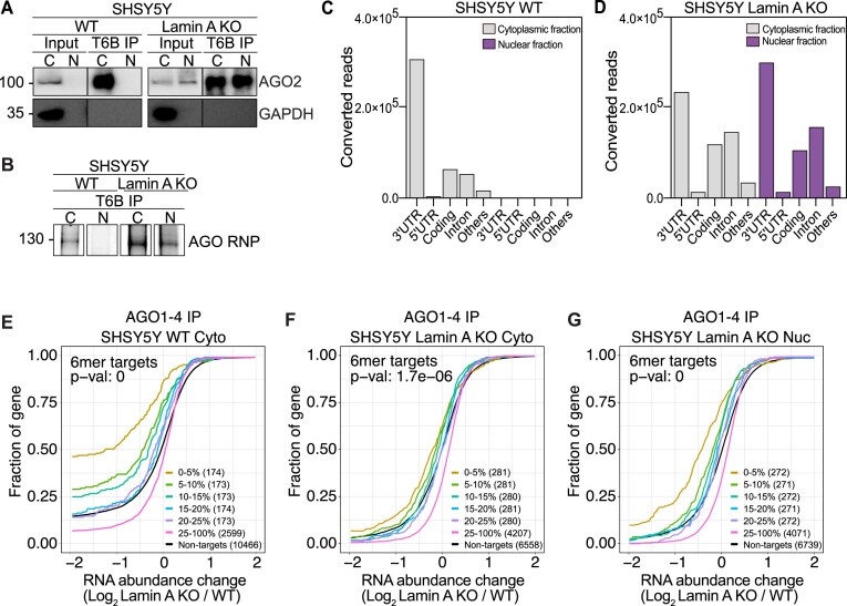
Figure 4.
Nuclear RNAi is diminished upon Lamin A KO. (A) Representative images of AGO2 and GAPDH immunoblots from SHSY5Y WT and Lamin A KO cells after immunoprecipitation of AGO1-4 using the T6B peptide. (B) Fluorescence SDS-PAGE of 3′ labeled AGO1-4 RNP in WT and Lamin A KO SHSY5Y cells. Distribution of AGO fPAR-CLIP sequence reads across target RNAs in SHSY5Y (C) WT cytoplasmic and nuclear lysate and (D) Lamin A KO cytoplasmic and nuclear lysate. Cumulative distribution of abundance changes in RNA in SHSY5Y (E) WT cytoplasmic fraction (F) Lamin A KO cytoplasmic fraction and (G) Lamin A KO nuclear fraction. For cumulative distribution assays the targets were ranked by number of binding sites from top 5–20% and 25–100% and compared to non-targets. WT, wild-type cells, Lamin A KO, Lamin A knock out cells; C and Cyto, cytoplasmic fraction; N and Nuc, nuclear fraction; IP, immunoprecipitation; fPAR-CLIP, fluorescent photoactivatable ribonucleoside-enhanced crosslinking and immunoprecipitation; RNP, ribonucleoprotein complex.

