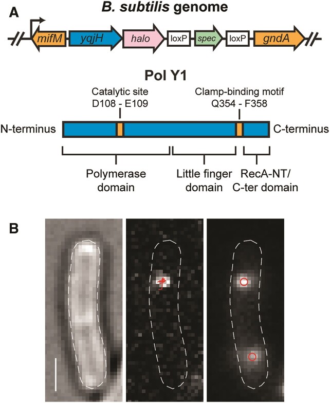
Figure 1.
(A) Cartoons of (top) Pol Y1-Halo (yqjH-halo) fusion and context in B. subtilis genome and (bottom) Pol Y1 domain organization and key residues. (B) Representative micrographs recorded with 13.9 ms integration time. Left: transmitted white light micrograph of B. subtilis cell with overlaid cell outline and 1 μm scale bar. Middle: fluorescence micrograph of single Pol Y1-Halo-JFX554 molecule with overlaid trajectory. Right: fluorescence micrograph of DnaX-mYPet foci with overlaid centroids.

