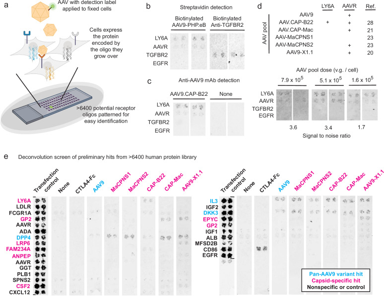
Fig. 1. High-throughput screen identifies AAV-binding human proteins.
a Schematic of AAV cell microarray screen. DNA oligos that encode individual membrane proteins are chemically coupled to slides in a known pattern, reverse transfecting the cells that grow on them and thereby creating spots of cells overexpressing a particular, known protein. Each protein is expressed in duplicate at two different locations on the slide. When AAVs are applied to the slides, enhanced binding can be detected from duplicate cell spots overexpressing cognate AAV receptors. b Known AAV capsid receptor interactions, such as AAVR and LY6A with AAV-PHP.eB, were used to optimize conditions for streptavidin-based detection of biotinylated capsids with two sets of replicate spots. Anti-TGFBR2 antibody was used as a non-AAV positive control. Uncropped blots in source data. c AAVR and LY6A interaction with AAV9.CAP-B22 were used to optimize conditions for anti-AAV9 antibody direct detection of unmodified capsids with two sets of replicate spots. Anti-TGFBR2 antibody was used as a non-AAV control. Uncropped blots in source data. d Pooled AAV capsid screening conditions were optimized by varying the concentrations of individual capsids within the pool to maximize signal to noise after direct detection with anti-AAV9 antibody, with two sets of replicate spots. v.g.: viral genomes. Uncropped blots in source data. e Pooled screening identified preliminary hits which were deconvoluted by individual-capsid screens, identifying previously-unreported potential capsid-binding proteins by direct detection with anti-AAV9 antibody. Transfection control condition detected fluorescent protein reverse transfected along with each receptor. None condition was treated only with anti-AAV9 antibody. Proteins in cyan were identified in all individual AAV screens, and likely represent interactions outside the engineered regions of AAV9. Proteins in magenta specifically bind to at least one engineered capsid. Uncropped blots in source data. Panel a created with BioRender.com released under a Creative Commons Attribution-NonCommercial-NoDerivs 4.0 International license.

