. 2024 Sep 6;12(9):e009603. doi: 10.1136/jitc-2024-009603

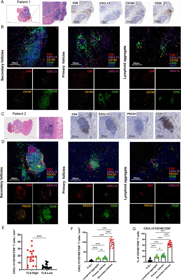
Figure 2. CXCL13+ CD160+ CD8+ T cells and CXCL13+ PDCD1+ CD4+ T cell around TLSs at different stages of maturation were assessed by multiplex immunofluorescence. (A) Resected gastric tissues stained with H&E or subjected to immunohistochemistry (IHC) detection for CD8, CXCL13, CD160 and CD20. The framed areas are shown adjacently at a higher magnification. Images were captured under a light microscope. Scale bar, 500 µm. (B) Multiplex immunofluorescence staining of human gastric cancer tissues for detection of CXCL13 (pink), CD160 (orange), CD8 (red), CD20 (green) and DAPI in gastric cancer tissues. Scale bar, 200 µm. (C) Tumor sections stained with H&E and IHC, including CD4, CXCL13, PDCD1 and CD20. The framed areas are shown adjacently at a higher magnification. Scale bar, 500 µm. (D) Multiplex immunofluorescence experiments were performed to detect the degree of CXCL13+ PDCD1+ CD4+ T-cell infiltration around TLSs at different stages of maturation. Antibody panel: CXCL13 (pink), PDCD1 (orange), CD4 (red), CD20 (green). Scale bar, 200 µm. (E) The numbers of CXCL13+ CD160+ CD8+ T cells in patients with low (n=17) versus high (n=17) levels of TLSs were compared. (F) The density of CXCL13+ CD160+ CD8+ T cells were higher around mature TLSs. (G) Percentage of CXCL13+ CD160+ CD8+ T cells among CD160+ CD8+ T cells around TLSs at different stages of maturation. Data are presented as the mean±SD. ns, not significant. *p<0.05, ***p<0.001, two-tailed Student’s t-test. DAPI, 4’, 6-diamidino-2-phenylindole; TLSs, tertiary lymphoid structures.

Figure 2