. 2024 Sep 6;12(9):e009603. doi: 10.1136/jitc-2024-009603

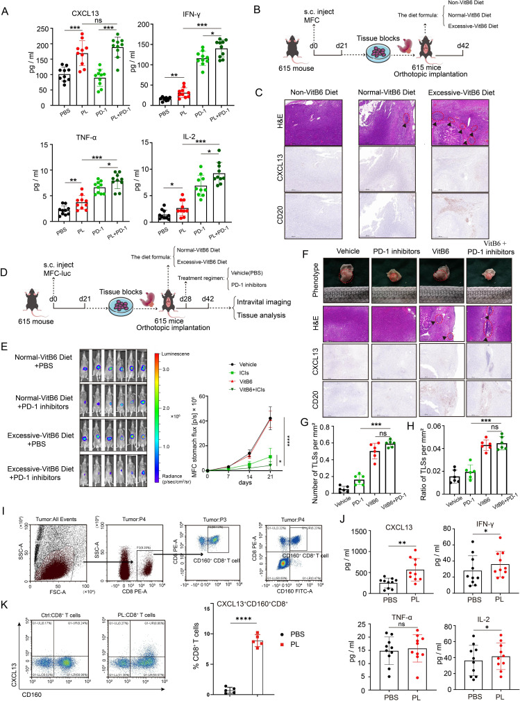
Figure 6. Vitamin B6 could promote the expression and secretion of CXCL13 in CD160+ CD8+ T cells. (A) Quantification of cytokine/chemokine including CXCL13, IL-2, TNF-α and IFN-γ, in PDTFs in the presence of different drugs measured by ELISA. (B) The schematic diagram of orthotopic transplanted tumor model with diets containing various amounts of vitamin B6 (n=6, each group). (C) Mouse orthotopic stomach xenograft tumor tissues stained with H&E and IHC detection for CD20 and CXCL13. Scale bar, 500 µm. (D) The schematic diagram of orthotopic transplanted tumor model fed with different drugs or diets (n=6, each group). (E) The representative images of mouse bioluminescence imaging at week 3 (left panel) and the corresponding quantification analysis (right panel). (F) Representative micrographs of xenografts stained with H&E and IHC detection for CD20 and CXCL13. Scale bar, 500 µm. (G–H) Density of TLSs (left panel) and ratio of tumor area occupied by TLSs (right panel). (I) Gating strategy for CD160+ CD8+ T cells. (J) Quantification of cytokine/chemokine including CXCL13, IL-2, TNF-α and IFN-γ, in supernatants from CD160+ CD8+ T-cell cultures in the presence or absence of PL measured by ELISA. (K) Flow cytometric analysis and corresponding quantification of CXCL13+ CD160+ CD8+ T cells with or without PL treatment. Data are presented as the mean±SD. ns, not significant. *p<0.05, **p<0.01, ***p<0.001, ****p<0.0001, two-tailed Student’s t-test. ICIs, immune checkpoint inhibitors; IFN, interferon; IHC, immunohistochemistry; IL, interleukin; MFC, mouse forestomach carcinoma; PBS, phosphate-buffered saline; PL, pyridoxol; PD-1, programmed cell death protein 1; s.c, subcutaneous injections; TLS, tertiary lymphoid structures; TNF, tumor necrosis factor.

Figure 6