Fig. 4.
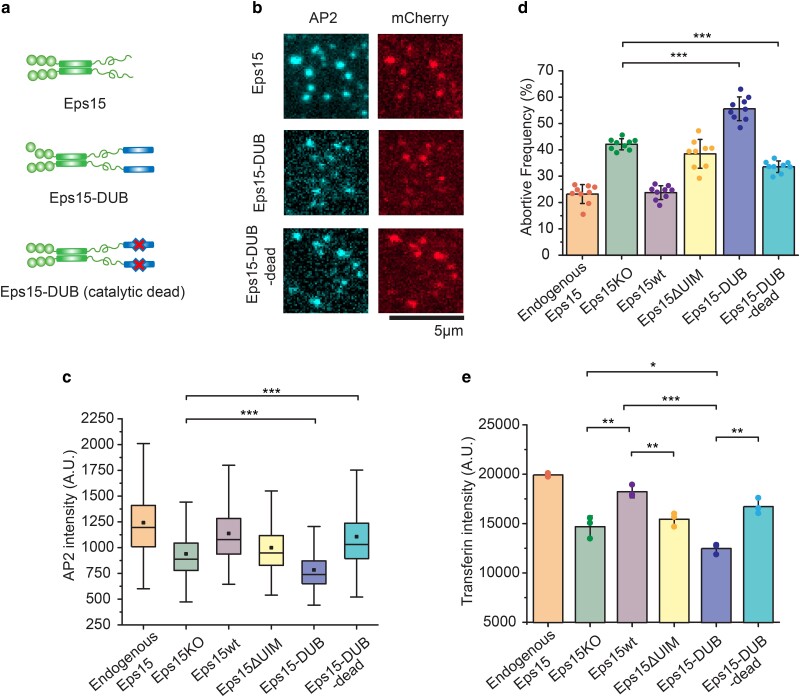
Fusion of a deubiquitinating enzyme to Eps15 results in even more short-lived endocytic events than deletion of Eps15. a) Schematic of Eps15 dimeric form, Eps15-DUB (DUB fused to C-terminal end of Eps15), and Eps15-DUB-dead (same with DUB but with a mutation that makes DUB catalytically dead). mCherry is not shown in the cartoon but all three constructs have mCherry at their C terminus for visualization. b) Representative images showing Eps15, Eps15-DUB, and Eps15-DUB-dead colocalization with AP2 in endocytic sites when Eps15KO SUM cells were transfected to express corresponding proteins. Scale bar = 5 μm. c, d) Box plot of endocytic pits AP2 intensity (c), and frequency of short-lived structures (d) under each condition. Endogenous Eps15, Eps15KO, Eps15wt, and Eps15ΔUIM are adopted from the same data shown in Figure 2. For Eps15-DUB, n = 9 biologically independent cell samples were collected and in total 8,640 pits were analyzed. For Eps15-DUB-dead, n = 9 and 7,420 pits were analyzed. Dots represent the frequency from each sample. e) Transferrin uptake under each condition measured by flow cytometry. N = 3 independent samples were measured for each group. An unpaired, two-tailed Student's t-test was used for statistical significance. *P < 0.05, **P < 0.01, and ***P < 0.001. Error bars represent standard deviation. Cells were imaged at 37°C for all conditions.

