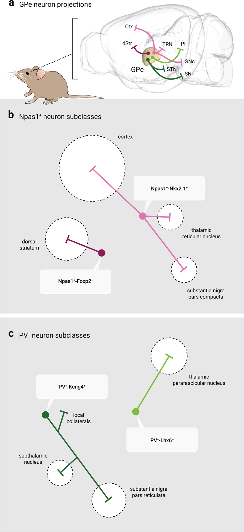
Figure 4. Distinct efferent projections of GPe neuron subtypes.

a. Map displaying the major projection targets of PV+ neurons and Npas1+ neurons. Note that GPe neuron projections are widespread and extend beyond the traditional basal ganglia nuclei, in contrast to the classic indirect pathway model. Magenta: Npas1+-Foxp2+ neurons; Pink: Npas1+-Nkx2.1+ neurons; Dark green: PV+-Kcng4+ neurons; Light green: PV+-Lhx6− neurons. Abbreviations: Ctx, cortex; dStr, dorsal striatum; Pf, thalamic parafascicular nucleus; SNr, substantia nigra pars reticulata; SNc, substantia nigra pars compacta; STN, subthalamic nucleus; TRN, thalamic reticular nucleus.
b. Schematic of the projection targets of Npas1+ neuron subtypes. Npas1+-Foxp2+ neurons represent 60% of the total Npas1+ neuron class and project exclusively to the dorsal striatum, targeting spiny projection neurons. Npas1+-Nkx2.1+ neurons represent 40% of the total Npas1+ neuron class and project to the cortex, thalamic reticular nucleus, and substantia nigra pars compacta.
c. Schematic of the projection targets of identified PV+ neuron subtypes. PV+-Kcng4+ neurons overlap with canonical PV+ neurons; they project to the subthalamic nucleus, substantia nigra pars reticulata and provide collateral inhibition to local GPe neurons. PV+-Lhx6− neurons project to the thalamic parafascicular nucleus. Size of the target areas (dotted circles) in b, c are artistic renderings based on the volume of the target areas and do not reflect the axonal density, synaptic strength, or contacts formed by GPe neuron subclasses.
