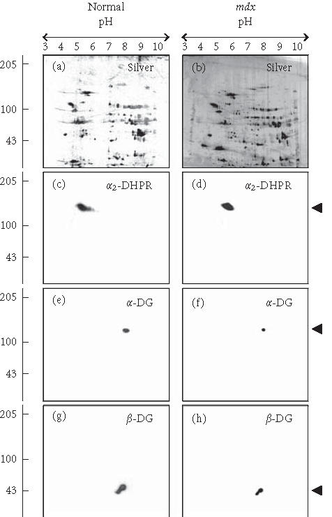
Figure 2.

Reduced expression of the dystroglycan complex in dystrophic heart muscle. Shown are silver-stained gels ((a), (b)) and identical immunoblots ((c), (d), (e), (f), (g), and (h)) of 24-week-old normal ((a), (c), (e), (g)) and age-matched mdx ((b), (d), (f), (h)) total heart extracts. Immunoblots were labelled with antibodies to the α2-subunit of the dihydropyridine receptor (α2-DHPR) ((c), (d)), α-dystroglycan (α-DG) ((e), (f)), and β-dystroglycan (β-DG) ((g), (h)). The position of immuno-decorated spots is marked by arrow heads. The pH values of the first-dimensional gel system and molecular mass standards (in kd) of the second dimension are indicated on the top and on the left of the panels, respectively.
