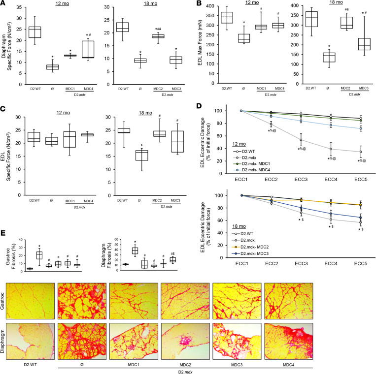
Figure 3. Microdystrophin provides partial rescue of D2.mdx skeletal muscle.
Male D2.mdx mice were treated with microdystrophin (μDys) gene therapy at 1 month of age (refer to Figure 2A). (A–D) At the terminal endpoints of 12 and 18 months, ex vivo muscle function was performed for the diaphragm (A) and extensor digitorum longus muscles (EDL) (B–D) of D2.WT, untreated D2.mdx, and μDys-treated D2.mdx mice (n = 6–10). (E) Representative PSR-stained images of the gastrocnemius and diaphragm muscles with accompanying fibrosis quantifications for these groups. Scale bar: 75μm. Data were analyzed using 1-way ANOVA with Tukey HSD post hoc tests (α = 0.05) and displayed as box-and-whisker plots (A–C and E) (boxes indicate second and third quartiles, and error bars represent the minimum and maximum values) or mean ± SEM. *P < 0.05 compared with WT; #P < 0.05 compared with untreated D2.mdx; %P < 0.05 compared with MDC1; $P < 0.05 compared with MDC2; &P < 0.05 compared with MDC3; @P < 0.05 compared with MDC4.

