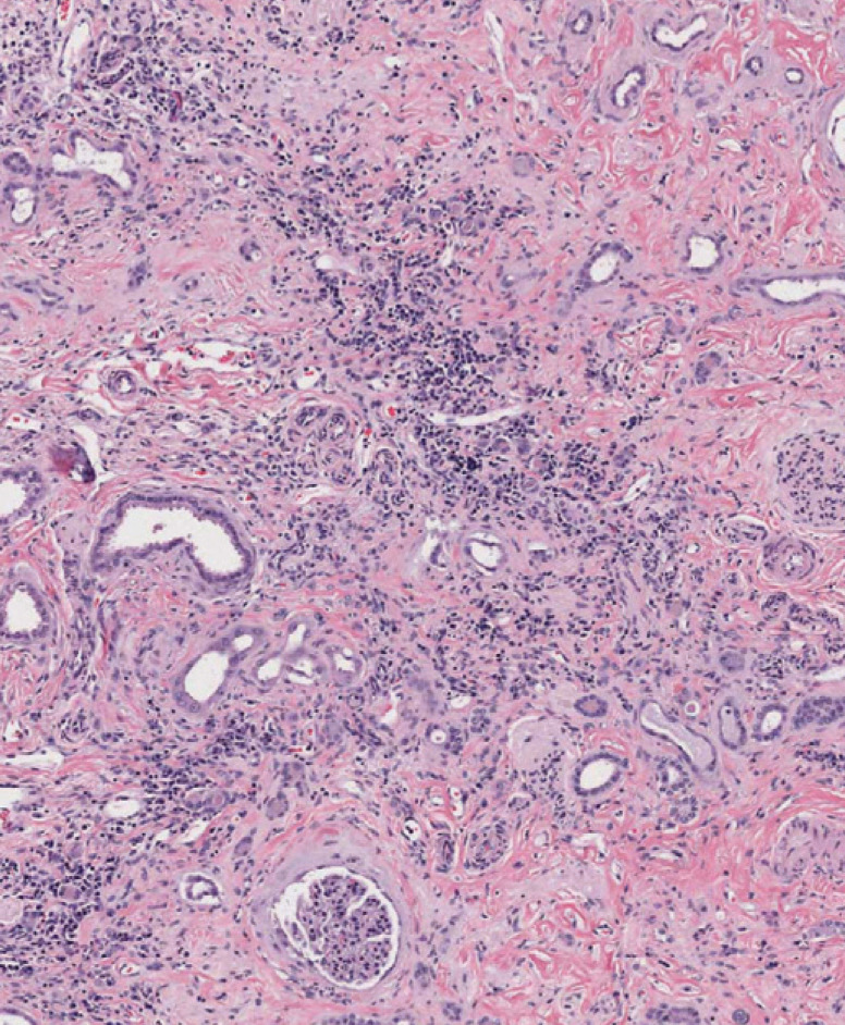
FIG 2.

Advanced histopathologic lesion of CKD. Note the large amount of fibrous tissue, loss of nephron mass (tubules and glomeruli), and interstitial inflammation with lymphoplasmacytes. Some tubules are dilated, presumably due to obstruction further down the nephron. Hematoxylin and eosin. Magnification × 8
