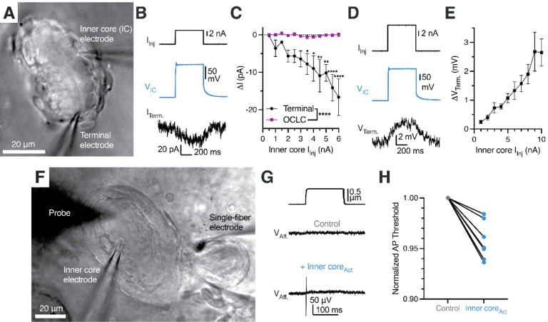
Figure 7. Activation of lamellar Schwann cells reduces the mechanosensitivity threshold of the Pacinian corpuscle.
(A) Bright-field image of simultaneous paired patch clamp recordings from one LSC and an associated Pacinian afferent of the same corpuscle.
(B) Exemplar traces showing current injection stimulus applied to a LSC (top), voltage response of the LSC (middle) and current response of the afferent terminal voltage-clamped at −60 mV (bottom).
(C) Quantification of current responses in the afferent terminal and an OCLC upon current injection into an LSC. Data shown as mean ± SEM from 4 afferent terminal and 4 OCLCs recordings. Statistics: two-way ANOVA with Holm-Šidák post-hoc tests (*p<0.05, **p<0.01, ****p<0.0001)
(D) Exemplar traces showing a current injection stimulus applied to a LSC (top), voltage response of the LSC (middle) and voltage response of the current-clamped afferent terminal (bottom).
(E) Quantification of voltage response in the afferent terminal upon current injection into an LSC. Data shown as mean ± SEM from 7 recordings.
(F) Bright-field image of simultaneous paired patch clamp recordings from one LSC and single-fiber recording of an associated Pacinian afferent of the same corpuscle while applied mechanical stimuli with the marked probe to measure AP threshold.
(G) Mechanical stimulus (top) and single-fiber recordings of the Pacinian afferent during the absence (middle) or presence (bottom) of LSC-inner core activation via 6 nA current injection
(H) Quantification of the effect of LSC activation by depolarizing current injection (6 nA) on the threshold of mechanical activation in Pacinian afferent evoked by to a square indentation step. Lines connect data from individual paired recordings. Mechanical threshold during inner core activation was lower than the normalized control threshold (p = 0.001, one sample t-test).

